DAN encourages divers and dive operators to comply with federal and state social distancing orders and to stay up to date on the recommendations of the CDC, WHO and your local health department. We acknowledge that some people will continue to dive, and we encourage them to take steps to lower their risk of disease transmission and minimize the risk of injury to avoid further stressing hospitals and emergency medical services.
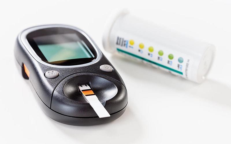
Diabetes is a major chronic disease that affects millions of people worldwide with an increasing trend. In the United States, more than 14 percent of adults are affected. Insulin-dependent diabetes mellitus (IDDM) affects up to half a million people of all ages, out of which 150,000 are younger than 19 years old. Many people continue to be productive members of the community and pursue various interests and careers despite having diabetes. However, when it comes to diving, the diving medicine community has long maintained the conservative position that IDDM is an absolute contraindication for diving. Recognizing that a substantial number of divers are diving successfully (either openly or surreptitiously) with diabetes in spite of the restriction has led many to believe that it is time to acknowledge this fact and re-examine the position concerning diabetes in diving.
A workshop addressing the issues of diabetes and recreational diving was jointly sponsored by the Undersea and Hyperbaric Medical Society (UHMS) and Divers Alert Network (DAN) on June 19, 2005 in Las Vegas, Nevada. They brought together experts and interested parties from within and beyond the international diving community. At the workshop, participants reviewed the existing data, discussed concerns, and finally developed consensus guidelines to address diabetes and recreational diving. The issues concerning professional diving require future, separate deliberations.
The consensus guidelines were released with the clear statement that it is a set of guidelines, not rules and with an understanding that various interest groups must have the flexibility to use the guidelines as they best serve their community’s needs.
This consensus reflects a more inclusive approach and provides guidelines on how to individually evaluate fitness to dive and how to keep it safe for those who qualify. Not everybody with diabetes who wishes to dive will be able to do so; there are various conditions and states of diabetes that would make diving with the condition too risky for divers and for those diving with them.
The guidelines are designed for individual divers who are primarily responsible for their own health and safety. They should adhere to these guidelines developed to improve their protection and that of their dive partners. The guidelines aim to also assist primary physicians and diving physicians evaluating and monitoring divers with diabetes. Other divers should be aware of the guidelines too, and be mindful of special considerations when buddied or leading dives with divers with diabetes.
Who may qualify for recreational scuba diving and how should they be monitored?
Individuals with diabetes who wish to dive must undergo the same medical fitness evaluation as other candidates to ensure first, that no other exclusionary conditions (e.g., epilepsy, pulmonary disease, heart disease, etc.) exist; and second, that there are no complications of diabetes that may increase the risk of injury while diving.
They should be 18 years or older (≥16 years if in special training program), with a well-established treatment, well maintained plasma glucose level and the ability to sustain those levels efficiently in the course of changing demands of daily activities. Candidates and divers with diabetes have to undergo mandatory medical examination annually, and if over 40 years old, they should be regularly evaluated for silent cardiovascular disease.
How to dive with diabetes
Candidates who pass the fitness evaluation and master regular scuba training, must also learn and adhere to the diabetic diving protocol. They should dive only in comfortable environmental conditions, with no overhead. Their dive should not exceed the depth 30 meters of sea water (100 fsw), duration of one hour nor involve compulsory decompression stops.
Divers with diabetes should dive with a buddy who is informed of their condition and is aware of the appropriate response in the event of a hypoglycemic episode. It is recommended that the buddy does not have diabetes.
Glucose management on the day of diving
Divers with diabetes whose medication may put them at risk of hypoglycemia, should use a protocol to manage their health on the day of diving.
Divers with diabetes should carry oral glucose in a readily accessible and ingestible form at the surface and during all dives. It is strongly recommended that parenteral glucagon is available at the surface. The dive buddy or another person at the surface should be knowledgeable in the use of glucagon. If symptoms or indications of hypoglycemia are noticed underwater, the diver should surface, establish positive buoyancy, ingest glucose and leave the water. An informed buddy should be in a position to assist throughout this process. Use of an “L” signal with the thumb and index finger of either hand is recommended as a signal for suspected hypoglycemia.
Blood glucose levels should be checked at the end of every dive. Appropriate response to the measured level can be determined by the individual mindful of his or her plans for the rest of the day. It should be noted that the requirements for blood glucose status remain the same for any subsequent dive. In view of the recognized potential for late decrements in blood glucose levels following diving, it is strongly recommended that the level is checked frequently for 12-15 hours after diving.
Divers with diabetes are strongly recommended to pay particular attention to adequate hydration on days of diving. Elevated blood glucose will lead to increased diuresis. While the data are limited, there is some evidence from divers with diabetes that an increase in hematocrit observed post-dive (suggesting dehydration) can be avoided by deliberate ingestion of fluid.
Divers with diabetes should log all dives, associated diabetic interventions and results of all blood glucose level tests conducted in association with diving. This log can be used to refine future planning in relation to diving.
Guidelines for recreational diving with diabetes
Selection and Surveillance
Age ≥18 years (≥16 years if in special training program)
Delay diving after start/change in medication:
Three months with oral hypoglycemic agents (OHA)
One year after initiation of insulin therapy
No episodes of hypoglycemia or hyperglycemia requiring intervention from a third party for at least one year
No history of hypoglycemia unawareness
HbA1c ≤9% no more than one month prior to initial assessment and at each annual review
values >9% indicate the need for further evaluation and possible modification of therapy
No significant secondary complications from diabetes
Physician/Diabetologist should carry out annual review and determine that diver has good understanding of disease and effect of exercise
in consultation with an expert in diving medicine, as required
Evaluation for silent ischemia for candidates >40 years of age
after initial evaluation, periodic surveillance for silent ischemia can be in accordance with accepted local/national guidelines for the evaluation of diabetics
Candidate documents intent to follow protocol for divers with diabetes and to cease diving and seek medical review for any adverse events during diving possibly related to diabetes
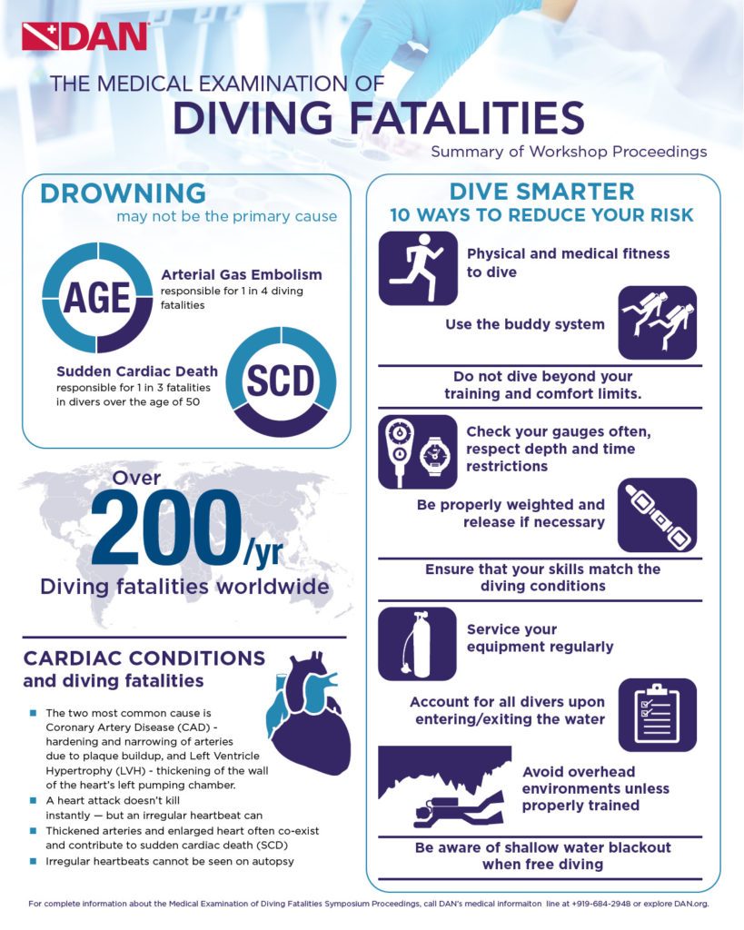
The DAN/ Undersea & Hyperbaric Medical Society (UHMS) sponsored Medical Examination of Diving Fatalities Symposium was held on June 18, 2014 in St. Louis, Missouri. Although the symposium was geared towards medical examiners, many of the issues discussed in the workshop are pertinent to dive professionals.
Why It May Not Be Drowning
A large number of deaths in scuba ascribed to drowning are in fact due to other causes: specifically, sudden cardiac death (SCD), and to a lesser extent, arterial gas embolism (AGE).
Some cases which have been labeled as “immersion” or “drowning” have subsequently been found to be due to other causes. Some of the more unusual causes include inhalation of inert gas (nitrogen), air hose entanglement (entrapment), and cuttlefish attack that caused perforated tympanic membrane, leading to panic, rapid ascent and gas embolism; there were also other causes labeled drowning.
Most medical examiners would call it drowning, simply because someone was in the water.
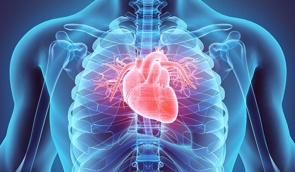
Cardiac Conditions Are Common Causes
Sudden cardiac death (SCD): two most common causes of SCD sudden causes in adults are coronary artery disease and left ventricle hypertrophy (LVH).
Atherosclerotic heart disease: it is not the heart attack that kills the person instantly, heart attacks and subsequent damage to the myocardium kill people over a time course of hours to days. It is the dysrhythmia that kills people instantly.
You can’t see an arrhythmia on autopsy.
Left Left ventricular hypertrophy (LVH): atherosclerotic disease often co-exists with another risk factor for SCD and that LVH. If you don’t recognize it, you are missing a huge risk factor for sudden death.
LVH may play a significant role in SCD in divers due to the stress on the body from diving may precipitate arrhythmias and death.
If we know what risk factors to look for, we may be able to improve our fitness to dive screenings and potentially prevent some of these deaths.
Looking for Preventable Causes of Death
Fatality investigation: in most cases investigation usually ends with establishing proximal cause of death. Unintentional or natural cause of death investigation usually stops short of pursuing root causes.
Injury research depends on the quality of data pro vided by investigation. Legal investigation may provide answers on questions of how it happened but often not concerned with “why”. The medical examination may answer what were the cause of death and the mode of death.
Field Investigations: Preserving the Evidence
Three general patterns to a diver’s death:
First, death occurs underwater with no rescue or resuscitation attempted. Disadvantage by possible delay in between when the diver dies and is recovered – autopsy info can be altered or affected.
Second, diver has a triggered even in the water and is brought to shore or boat for attempted rescue but dies prior to transportation to medical facility. Usually provides a witness to describe what happened.
Third, diver is transported to a medical facility and survives for a few hours or days. Advantages are that imaging and lab tests may guide determination of the cause of death, however autopsy findings may be altered by the survival interval and medical intervention.
Diving conditions and diving equipment may cause or contribute to a diver’s death. Information may be lost as witnesses leave, forget equipment, or worse, equipment is returned to the family.
Field investigation is categorized into six parts:
History
Antemortem events
The environment
Body recovery
Medical care administered before death
Body and equipment recovery and documentation and preservation of evidence
Post Mortem: How-To
Very few forensic pathologists have significant experience with the investigation of fatalities involving divers who were breathing compressed gas.
Fewer than 100 combined deaths occurring in the US, Canada, and the Caribbean each year.
Pathologists should be aware of the circumstances surrounding the fatal dive mishap, but the diver’s past medical and surgical history, recent health status, and any medications taken on a regular basis and on the day of the mishap need to be known.
Cardiovascular disease in particular is a frequent factor in a diving related fatality, especially in older divers.
What Medical Examiners Need to Know About Rebreathers
Three main root causes of fatal accidents with rebreathers:
Diver error (the most common)
Mechanical problems
Electronic problems
An autopsy cannot reveal hypoxia, hyperoxia, or hypercapnia (the three most common causes of rebreather fatalities). In most cases, the medical examiner cannot detect the root cause of a rebreather fatality.
Expert Panel Review of Investigation and Autopsy Findings
Guidelines identified by common trends seen in diver death:
Ensure physical fitness to dive: train for your sport and be sure that you exercise regularly and follow a healthy diet.
Use the buddy system.
Follow your training: check your gauges often, respect depth and time restrictions, and do not dive beyond your training limits.
Weight yourself properly and remember to release your weights when appropriate.
Ensure that your skill level and familiarity are appropriate for conditions.
Have your equipment serviced and maintained regularly.
Account for all divers (a physical, individual response should be received from every diver before entering/after exiting).
Avoid overhead environments unless properly trained and equipped.
Breath-hold divers should remember to use the buddy system and be aware of the dangers of shallow-water blackout.
Team of doctors cooperating while reading medical chart. Focus is on background, on two young colleagues.
Appendix F in the Recreational Diving Fatalities Workshop Proceedings is the Autopsy Protocol for Recreational Diving Fatalities by Dr. James Caruso
Vann RD, Lang MA, eds. Recreational Diving Fatalities. Proceedings of the Divers Alert Network 2010 April 8-10 Workshop. Durham, NC: Divers Alert Network, 2011. IBSN#978-0615-54812-8.
History
This is absolutely the most important part of the evaluation of a recreational diving fatality. Ideally, one should obtain significant past medical history with a special focus on cardiovascular disease, seizure disorder, diabetes, asthma and chronic obstructive pulmonary disease. Medications taken on a regular basis as well as on the day of the dive should be recorded, and information regarding how the diver felt prior to the dive should be obtained. Any history of drug or alcohol use must also be noted.
The dive history is extremely important. If possible, the investigator should find out the diver’s experience and certification level. The most important part of the history will be the specific events related to the dive itself. The dive profile (depth, bottom time) is an essential piece of information, and if the diver was not diving alone, eyewitness accounts will be invaluable. With the near-universal use of dive computers, the computer used by the deceased diver should be interrogated, and if it has a download function all recent dives should be reviewed.
Not only will the last dive or dive series be invaluable to the investigation, much can be learned about the diver by looking at previous dives made, including frequency, depth, ascent habits and with certain computers even breathing gas usage. Written dive logs are also a valuable source of information related to the diver’s experience level and dive habits.
Questions Include:
When did the diver begin to have a problem (predive, descent, bottom, ascent, postdive)?
Did the diver ascend rapidly (a factor in air embolism and pulmonary barotrauma)?
Was there a history of entrapment, entanglement or trauma?
If resuscitation was attempted, what was done, and how did the diver respond?
External Examination and Preparation
A thorough external examination including documentation of signs of trauma or animal bites or envenomation should be carried out. Palpate the area between the clavicles and the angles of the jaw for evidence of subcutaneous emphysema. X-rays of the head, neck, thorax and abdomen should be taken to look for free air. Postmortem CT imaging can be obtained as an alternative.
Modify the initial incision over the chest to make a “tent” or “pocket” out of the soft tissue (an “I” shaped incision) and fill this area with water. A large bore needle can be inserted into the second intercostal spaces on each side; if desired, any escaping air can be captured in an inverted, water-filled, graduated cylinder for measurement and analysis. As the breast-plate is removed, note any gas escaping from vessels. An alternative test for pneumothorax consists of teasing through the intercostal muscles with a scalpel and observing the relationship between the visceral and parietal pleura as each pleural cavity is entered. If the two pleural layers are still adjacent until the pleural cavity is breached, there is no evidence of a pneumothorax. If a pneumothorax had occurred during the final dive, the lung would already be at least partially deflated and not up against the parietal pleura.
The pericardial sac can be filled with water and the chambers of the heart may be incised with a scalpel to look for any intracardiac gas. As was possible for the pleural cavities, escaping gas may be captured and analyzed, but most medical examiner offices do not have the resources for such endeavors. After the mediastinum, heart and great vessels have been examined under water for the presence of gas, the water may be evacuated and a standard autopsy may be performed.
Carefully examine the lungs for bullae, emphysematous blebs and hemorrhage.
Note any interatrial or interventricular septal defects. Carefully check for evidence of cardiovascular disease and any changes that would compromise cardiac function.
Toxicology: Obtain blood, urine, vitreous, bile, liver and stomach contents. Not all specimens need to be run, but at least look for drugs or abuse. If an electrolyte abnormality is suspected or if the decedent is a diabetic, the vitreous fluid may prove useful for analysis.
Prior to opening the skull, tie off all the vessels in the neck to prevent artifactual air from entering the intracranial vessels. Tie the vessels at the base of the brain once the skull is opened. Disregard bubbles in the superficial veins or venous sinuses. Examine the meningeal vessels and the superficial cortical vessels for the presence of gas. Carefully examine the Circle of Willis and middle cerebral arteries for bubbles.
Have an expert evaluate the dive gear. Are the cylinders empty? If not, the gas should be analyzed for purity (a little carbon monoxide goes a long way at depth). All gear should be in good working order with accurate functioning gauges.
Possible Findings
Air Embolism
Intra-arterial and intra-arteriolar air bubbles in the brain and meningeal vessels, petechial hemorrhages in gray and white matter, evidence of COPD or pulmonary barotrauma (pneumothorax, pneumomediastinum, subcutaneous emphysema), signs of acute right heart failure, pneumopericardium, air in coronary and retinal arteries.
Carbon Monoxide Poisoning
Deaths due to carbon monoxide poisoning are rare in recreational diving, but they do occur. Autopsy findings are similar to carbon-monoxide-related deaths in other settings, with the classic finding of a cherry red color to the organs and blood. A carboxy-hemoglobin measurement should be obtained as routine toxicology in all diving- related deaths to exclude the contribution of contaminated breathing gas.
Decompression Sickness
Lesions in the white matter in the middle third of the spinal cord including stasis infarction, if there is a patent foramen ovale (or other potential right to left heart shunt) a paradoxical air embolism can occur due to significant venous bubbles entering the arterial circulation.
Drowning
While drowning essentially remains a diagnosis of exclusion, there are some anatomic findings that are observed with considerable frequency. The lungs usually appear hyperinflated and can even meet at the midline when the anterior chest wall is removed. Lungs are typically heavy and edematous, and pleural effusions may be present. A moderate amount of water and even some plant material may be present, not only in the airway but also in the esophagus and stomach. Dilatation of the right ventricle of the heart is commonly observed as is engorgement of the large central veins. Fluid is also often found in the sphenoid sinus.
Venomous Stings or Bites
A bite or sting on any part of the body, unexplained edema on any part of the body, evidence of anaphylaxis or other severe allergic reaction.
Interpretation
The presence of gas in any organ or vessel observed at the autopsy of someone who breathed compressed gas just prior to death is not conclusive evidence of decompression sickness or air embolism. During a dive, especially one of considerable depth or bottom time, inert gas dissolves in the tissues, and the gas will come out of solution when the body returns to atmospheric pressure. This, combined with postmortem gas production, will produce bubbles in tissue and vessels. The phenomenon has caused many experienced pathologists to erroneously conclude that a death occurred due to decompression sickness or air embolism.
Intravascular bubbles present predominantly in arteries and observed during an autopsy performed soon after the death occurred is suspicious for air embolism. The dive history will help support or refute this theory.
Gas present only in the left ventricle or if analysis shows the gas in the left ventricle has a higher oxygen content than that present on the right side would also be supportive for the occurrence of an air embolism.
Intravascular gas from decomposition or off-gassing from the dive would contain little oxygen and be made up of mostly nitrogen and carbon dioxide.
Deeper, longer dives can cause decompression sickness and significant intravascular (mostly venous) gas. Decompression sickness is rarely fatal and more commonly causes significant morbidity (illness and injury) in severe cases. Rapid ascents and pulmonary barotrauma are associated with air embolism.
Diving Fatalities Infographic
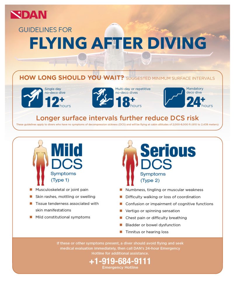
Guidelines for Flying After Diving
Proceedings Summary | DAN Flying After Diving Workshop
This workshop on flying after recreational diving was organized by Divers Alert Network (DAN) to bring together representatives from the recreational diving industry with experts from other diving communities. The workshop had two purposes: (a) to review the guidelines and experimental data developed since the first flying after diving workshop in 1989; and (b) to debate a consensus for new flying after recreational diving guidelines.
Previous consensus advised to wait 12 hours after a single no-stop dive, 24 hours after multi-day repetitive dive, and 48 hours after dives that required decompression stops. This was considered overly conservative. Subsequently, DAN proposed a simpler 24-hour wait after any and all recreational diving. There were objections to this on the grounds that the decompression sickness (DCS) risks of flying after diving (FAD) were too low to warrant such a long delay and would result in lost business for island diving resorts.
DAN flying after diving trials
Because little human experimental data could be found that was relevant to flying after recreational diving, DAN funded a series of trials at the Duke University Center for Hyperbaric Medicine and Environmental Physiology that were conducted from 1992-1999. Dry, resting volunteers tested nine single and repetitive dive profiles that were near the recreational diving no-decompression limits. The dives were followed by four-hour simulated flights at 8,000 feet (2,438 meters). In 802 trials, there were 40 DCS incidents during or after flight. For single no-stop dives to 60 fsw (feet of sea water; 18 msw, or meters of sea water) or deeper, there was no DCS for surface intervals of 11 hours or longer. For repetitive, no-stop dives, DCS occurred for surface intervals of less than 17 hours. The results of the study were used by the US Navy in 1999 to revise its rules for ascent to altitude following air diving. The new procedures were based on the diver’s repetitive group upon surfacing from a dive and on the expected post-dive altitude. While they were not formally tested in the laboratory prior to issue, no DCS cases have been reported to the Naval Safety Center to date. However, the number of times the new procedures have been used in the field was unknown.
Flying with DCS symptoms
The workshop reviewed recent FAD trials and available field data regarding flying after diving and flying with DCS symptoms. There were potentially important differences between field and chamber studies. Diving in the field involved immersion, exercise, and multiple days of diving, while the chamber trials occurred on a single day with dry resting divers. Thus, the chamber trials might not adequately simulate flying after diving as it actually occurs. As more divers fly with symptoms than develop symptoms during or after flight, flying with symptoms may be a greater health problem than symptoms that occur during or after flight. This is an educational issue, not a scientific issue. Divers need to be taught to seek medical advice rather than to fly if they note signs and symptoms consistent with decompression illness.
Diving nitrox and pre-breathing oxygen reduces risk of DCS in flying after diving
The benefits of oxygen pre-breathing after air diving were confirmed by trials conducted by the Special Operations Command (SOCOM). This organization was concerned with high-altitude parachute operations that might occur after air diving. Flying after diving trials were conducted with dry, resting divers who breathed air while exposed for 60 minutes at 60 fsw (18 msw). Dives were followed by simulated flights of two- or three-hour duration at an altitude of 25,000 feet (7,620 meters). It was demonstrated that this flight may cause DCS even without previous diving. When the dive was followed by a 24-hour surface interval and a three-hour flight, with divers breathing oxygen for 30 minutes immediately preceding flight, during ascent, and while at altitude there was no DCS in 23 trials. The study indicated that: (a) DCS risk was low for these flying after diving exposures, at least for dry resting divers; and (b) preflight oxygen might be an effective means for reducing DCS risk.
Considering possible impact of flying after diving rules on dive operations
One generally thinks of diving guidelines as based on medical safety, but safety is not the only yardstick humans use in establishing rules for living. Economics also has a major impact, albeit one not always articulated with comfort in the medical community. Economics was a primary issue in the 1991 discussion about the impact of DAN’s proposed 24-hour flying after diving guideline. Offshore diving operations felt they would needlessly lose business with a single 24-hour guideline. With this in mind, it was useful to approach the problem of flying after diving with an economic model in which the optimal preflight surface interval was determined by the economic interests of society as represented by divers, resorts, and insurers. Models of this nature depend on their assumptions, and no model can represent all situations, but economic modeling can differentiate between important and unimportant factors. In the model presented, for example, important factors included cost of a dive, number of days diving, aggressiveness of the dive and the DCS risk due to flying after diving. Unimportant factors included the probability of evacuation, the cost of treatment, the diver’s salary and the number of dives per day.
The consensus process
Science is a quantitative activity, while the determination of safety is a social process that considers the probability, severity and the costs of injury. Ultimately, the knowledgeable representatives of society make decisions about safety for society at large based on available information. The workshop participants were asked to reach consensus concerning:
a. whether flying after diving guidelines were needed for recreational diving; (b) whether the current guidelines were adequate; b. what the longest needed guideline might be; and c. if shorter guidelines were appropriate for short dives.
The ensuing discussion determined that guidelines were needed, and the evidence that had been presented demonstrated that existing guidelines were inadequate. After some debate it was decided that unless dive computers were used, written guidelines for recreational diving should be simple and unambiguous without the need for reference to tables such as the U.S. Navy procedures required. Three groups of divers were proposed for consideration:
a. uncertified individuals who took part in a “resort” or introductory scuba experience; b. certified divers who made an unlimited number of no-decompression air or nitrox dives over multiple days; and c. technical divers who made decompression dives or used helium breathing mixes.
Consensus recommendations for flying after diving
A minimum of 12-hour surface interval was recommended for the single no-decompression dive.
A minimum of 18-hour surface interval for multi-day repetitive diving.
Substantially longer than 18 hours after diving involving compulsory decompression, or using heliox and trimix.
Limitations
It was stressed that as the experimental trials described in the workshop had been conducted in a dry hyperbaric chamber with resting volunteers, longer guidelines might be needed for divers who were immersed and exercising. The effects of exercise and immersion on preflight surface intervals were seen to require experimental study. Additional studies were conducted since and the results will be published soon.
Vann RD. Executive Summary. In: Flying After Diving Workshop. Vann RD, ed. 2004. Durham: Divers Alert Network. ISBN 0-9673066-4-7. 16-19.
Flying After Diving Infographic
Guidelines for Patent Foramen Ovale and Diving
Proceedings Summary | DAN/UHMS PFO and Fitness to Dive Workshop
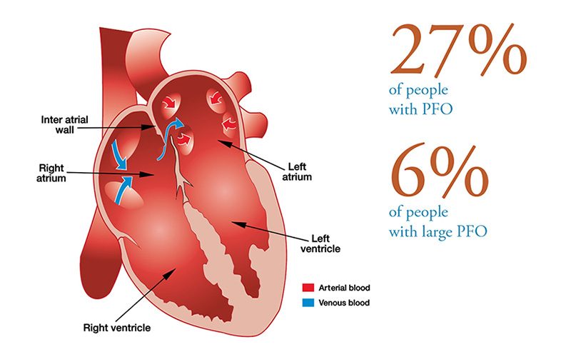
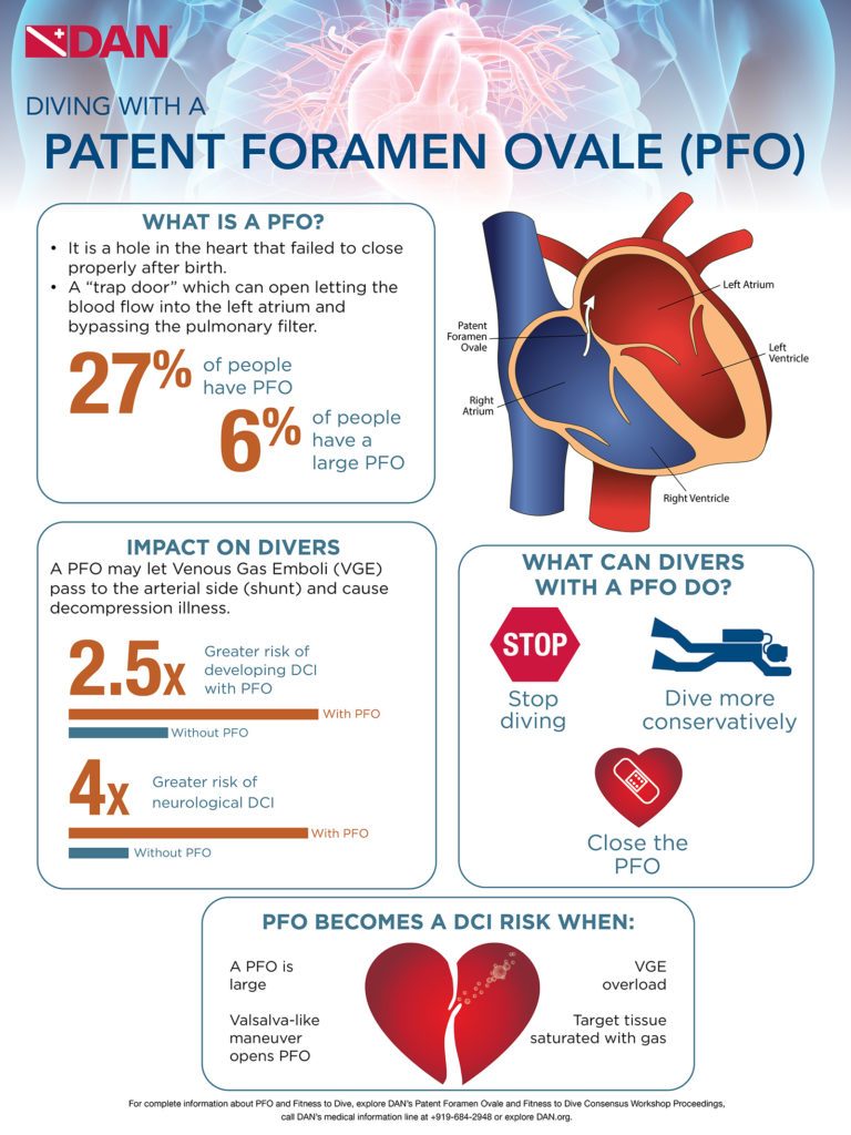
Prior to birth, oxygenated blood flows from the mother through the placenta to the heart of the fetus via the opening in the wall separating the left and right atrium (foramen ovale) into the fetal circulation. The foramen ovale has a “trap door” feature which opens due to the pressure of blood flow from the mother’s placenta entering the right atrium, and lets the blood pass to the left atrium. At birth, the lungs expand and the pressure in the left atrium increases and “slams shut” the foramen ovale. Shortly after birth the “door” fuses together, but in about 27 percent of people, it fails to fuse completely and results in a patent foramen ovale (PFO) also called persistent foramen ovale.
In people with PFO, if the pressure in the right atrium rises above the pressure in the left atrium, blood can flow from the right to the left atrium. The direct flow of blood from the right to the left atrium which bypasses the lungs is called right-to-left shunt (RLS). The RLS is known to let blood clots pass to the arterial side which can cause a stroke (brain trombo-embolism). Similarly, the PFO in divers may let gas bubbles from the venous blood — venous gas emboli (VGE) — pass the arterial side and cause decompression sickness.
Epidemiological studies have shown an association between PFO and certain types of neurological and cutaneous decompression sickness (DCS). The DCS risk in recreational divers has been reported at 3.6 cases per 10,000 dives, with 0.84 cases of neurological DCS per 10,000 dives and four-fold increase in risk with PFO. The overall risk of neurological DCS is low, even in the presence of a PFO. However, for some individuals, PFO seems to be a greater risk than predicted. Guidelines for PFO testing are aimed at identifying such individuals and managing their DCS risk.
The following guidelines were developed from the joint position statement on PFO and diving published by the South Pacific Underwater Medicine Society (SPUMS) the United Kingdom Sports Diving (UKSDMC), and the DAN sponsored workshop held in conjunction with the UHMS Annual Scientific Meeting in Montreal, Canada, June 2015.
Who Should Be Tested for PFO?
Routine screening for PFO at the time of dive medical fitness assessment (either initial or periodic) is not indicated. Consideration should be given to testing for PFO when there is a history of more than one episode of decompression sickness (DCS) with cerebral, spinal, vestibulocochlear or cutaneous manifestations.
Non-cutaneous manifestations of “mild DCI” as defined in the Remote DCI Workshop Proceedings [Consensus Statements, In: Management of Mild or Marginal Decompression Illness in Remote Locations, Workshop Proceedings (May 24-25, 2004). Mitchell SJ, Doolette DJ, Wachholz CJ, Vann RD, Eds. Divers Alert Network, Durham, NC, 2005, pp. 6-9.] are not indications for PFO investigation. Headache as an isolated symptom after diving is not an indication for PFO investigation.
PFO Testing and Evaluation Recommendations
PFO Testing
Testing is undertaken by centers well practiced in the technique.
The testing must include bubble contrast, ideally combined with trans-thoracic echocardiogram (TTE). Use of two-dimensional and color-flow echo cardiography without bubble contrast is not adequate.
The testing must include the use of provocation maneuvers to promote right-to-left shunt including Valsalva release or sniffing as described in the supporting references (both undertaken when the right atrium is densely opacified by bubble contrast).
What Does a Positive Test Mean?
A spontaneous shunt without provocation or a large, provoked shunt following diving when venous gas emboli are present is recognized as a risk factor for those forms of DCS with cerebral, spinal, vestibulocochlear or cutaneous manifestations.
Smaller shunts are associated with a lower but poorly defined risk of DCS. The significance of minor degrees of shunting needs to be interpreted in the clinical setting that led to testing.
Detection of a PFO after an episode of DCS does not guarantee that the PFO contributed to causation.
What Are the Options for Divers to Test Positive?
Following a diagnosis of PFO considered likely to be associated with increased DCS risk, the diver may consider the following options in consultation with a diving physician:
Stop diving.
Dive more conservatively. There are various strategies that might be employed to reduce the risk of significant venous bubble formation after diving, or the subsequent right-to-left shunting of such bubbles across a PFO. The appropriateness of this approach, and the strategies chosen, need to be considered on an individual basis, and in discussion with a diving medicine expert. Examples include: reducing dive times to well inside accepted no-stop limits; performing only one dive per day; use of nitrox with air dive planning tools; intentional lengthening of a safety stop or decompression time at shallow stops; avoidance of heavy exercise and unnecessary lifting or straining for at least three hours after diving.
Close the PFO. It is emphasized, however, that closing a PFO after an episode of DCS cannot be considered to provide assurance that DCS will not occur again. The options outlined above require careful consideration of the risks and benefits and the clinical setting that led to screening.
When Can Divers Who Undergo Closure Return to Diving?
Following closure of a PFO and before returning to diving, the diver requires a repeat bubble contrast echocardiogram demonstrating shunt closure, a minimum of three months after the closure. Diving should not be resumed until satisfactory closure of the PFO is confirmed, and the diver has ceased potent antiplatelet medication (aspirin is acceptable).
CAUTION Venous bubbles can also enter the systemic circulation through intrapulmonary shunts, although the role of this pathway in the pathogenesis of decompression sickness is not as well established as PFO. These shunts are normally closed at rest. They tend to open with exercise, hypoxia and beta adrenergic stimulation, and close with hyperoxia. It is therefore plausible that exercise, hypoxia and adrenergic stimulation after a dive could precipitate decompression sickness when it might not otherwise have occurred, while supplemental oxygen is likely to minimize this effect.
Facts About Divers With PFO
Divers with PFO have 2.5 times greater overall risk of DCS than divers without a PFO and four times greater risk of neurological DCS. However, the absolute incidence of neurological DCS in divers with PFO is estimated at 4.7 DCS cases per 10,000 dives.
A major study at the Mayo Clinic by Dr. Hagen and colleagues determined there is a large prevalence of PFO in young people, however it declines and levels off at approximately 27 percent. They also found that in each of the decade intervals, there is no difference in prevalence of PFOs between men and women.
Four studies were compared, determining the prevalence of RLS or large PFO in divers with spinal DCI is 44 percent compared to 14.2 percent in controls, those without prevalence of RLS or large PFO.
Half of the divers in the studies with RLS related DCI have a PFO that is a centimeter in diameter or larger, therefore the greatest risk of DCI is in those with the largest PFOs (six percent), not all divers with a PFO.
Cerebral, spinal, cutaneous and inner ear DCS have been associated with PFO, however the link between PFO and cutaneous and inner ear DCS is the strongest. In approximately 74 percent of cases present with isolate inner ear symptoms (no other symptoms of hyperbaric related issues), 80 percent of the cases had a large spontaneously shunting PFO.
There are factors necessary for PFO to contribute to DCS: you need to have a large PFO; venous gas emboli must form; bubble must cross the PFO (provocative factor to open PFO needed) to arterial circulation; and the bubbles must reach a target tissue while it is still supersaturated and vulnerable.