Metformin

Metformin and diabetes kit

Metformin is a widely used oral medication for the treatment of DM2 (Type 2 diabetes mellitus) and, in some cases, PCOS (polycystic ovary syndrome). While insulin helps lower blood sugar levels by helping cells absorb sugar from the blood, metformin lowers blood sugar by reducing sugar production in the liver and improving the body’s response to insulin.

Unlike many other diabetes medications, metformin does not stimulate insulin secretion, significantly reducing the risk of hypoglycemia (low blood sugar). In plain words, metformin alone doesn’t cause low blood sugars.

Metformin and Diving

Although metformin is generally well tolerated, divers using it must consider its effects under hyperbaric and immersion conditions. The key concerns for scuba divers include lactic acidosis, thermoregulation, dehydration, and hypoglycemia when combined with other medications for diabetes.

Risk of Lactic Acidosis

One rare but serious side effect of metformin is lactic acidosis — a condition in which lactic acid builds up in the bloodstream, leading to metabolic acidosis. While the overall incidence is low, diving presents several physiological stressors that could increase this risk, including:

  • Hypoxia (low oxygen levels)
  • Hypercapnia (high carbon dioxide levels)
  • Increased anaerobic metabolism from exertion (e.g., swimming against currents)
  • Impaired kidney function (which can reduce metformin clearance)

If anaerobic metabolism and carbon dioxide retention occurs, it may theoretically exacerbate lactate accumulation, especially in divers with impaired renal function.

Impaired Thermoregulation and Dehydration

Metformin reduces hepatic glucose output, impairing the body’s ability to produce heat under cold conditions. This effect, combined with the increased heat loss associated with immersion, may increase a diver’s susceptibility to hypothermia.

Additionally, metformin-associated gastrointestinal side effects (such as diarrhea or nausea) can contribute to dehydration, which is already a common concern in diving due to dry breathing gases and immersion diuresis (increased urine production underwater). Dehydration can impair circulation, slow nitrogen elimination, and increase decompression sickness risk.

Risk of Hypoglycemia

While metformin alone does not typically cause hypoglycemia, when combined with other glucose-lowering medications such as insulin or sulfonylureas, there is a risk of dangerously low blood sugar levels underwater. Symptoms of hypoglycemia—dizziness, confusion, weakness, and loss of consciousness—can be life-threatening in a diving environment.

Factors that could increase the risk of hypoglycemia while diving include:

  • Skipping meals before a dive
  • Extended dive times leading to prolonged fasting
  • Increased exertion consuming glucose faster than usual
  • Co-administration with insulin or sulfonylureas

Implications in Diving

For the Diver

  • Divers taking metformin should obtain medical clearance from a physician.
  • Monitor for trends in BGL. It is recommended to stay between 150-180mg/DL. Do not dive if your BGL is under 150mg/DL or over 300mg/DL
  • Diabetic individuals on metformin must monitor blood glucose 30 minutes before immersion and immediately after a dive.
  • Do not dive if hypoglycemic or if you’ve had a recent episode of hypoglycemia in the last 24 hours.
  • Individuals with renal impairment should consult a physician before diving to assess kidney function and metformin clearance.
  • Stay hydrated before, during, and after diving to reduce the risk of dehydration and lactic acidosis.
  • Avoid prolonged cold-water exposure, which can increase the risk of hypothermia and metabolic stress.
  • Divers taking metformin should obtain medical clearance from a physician.

For the Dive Operator

  • Staff should be trained in first aid and able to recognize possible medical emergencies.
  • Keep oral glucose or snacks on hand in case of post-dive blood sugar drops.

For the Physician

  • Consider evaluation of renal function and lactic acidosis risk.
  • Assess fitness for diving based on activity level.
  • If the diver takes additional diabetes medications (e.g., insulin, sulfonylureas), avoid extended or deep dives to mitigate hypoglycemia risks.

Mitral Valve Prolapse

Mitral Valve Prolapse Patient talking with her doctor during a cardiopulmonary stress test on a hospital

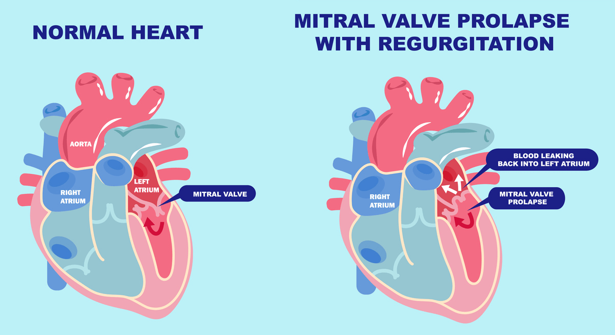
Mitral valve prolapse (MVP) is a condition in which the mitral valve — which allows blood to flow from the left atrium into the left ventricle — does not close properly, resulting in a backflow of blood called regurgitation. This can cause a characteristic murmur during auscultation. MVP is also known as “floppy valve syndrome” or “click-blow syndrome” and is more common in women.

The mitral valve has two leaflets. MVP is characterized by the expansion and contraction of one or both of these leaflets towards the left atria. If severe, then blood may pass or reflux and cause mitral reguritation. The causes of MVP are uncertain, but there are two forms: primary (usually hereditary) and secondary (associated with other diseases). Primary MVP may result from thick valves, irregular surfaces, or issues with tendon fibers. Secondary MVP is linked to additional diseases affecting blood flow, heart muscle function, or valve structure.

Doctors alternatively classify MVP as “flaccid valve syndrome” or “click-murmur syndrome.” This condition is common, particularly among women. MVP presents with a wide wide range of symptoms, which can include chest discomfort, an unexpected sensation in the chest during each beating, minimal or no symptoms, or even a myocardial infarction. Transitory loss of consciousness or a mild stroke (called “transitory ischemic attacks”) is another risk factor associated with MVP.

Understanding MVP

Understanding the potential risks for individuals with MVP who wish to scuba dive requires understanding the condition itself. MVP occurs when the mitral valve, which regulates blood flow between the left heart’s chambers, does not close properly. This faulty closure allows blood to leak backward into the left atrium, leading to symptoms like chest pain, fatigue, and heart palpitations. While MVP is typically benign, certain factors can increase the risk of complications: severe valve regurgitation, a family history of MVP-related complications, and an enlarged left atrium. Individuals with MVP should undergo regular cardiology check-ups to monitor their heart health and function.

Risks Associated with Diving with MVP

People with MVP should consider potential risks before scuba diving. The sport involves descending to depths where pressure increases significantly, placing additional strain on the heart and potentially causing complications. Individuals with MVP are at a higher risk of experiencing arrhythmias, abnormal heart rhythms, and the physical exertion involved in scuba diving can further increase the risk of arrhythmias.

Precautions for Individuals with MVP

If you have MVP and are considering diving, take certain precautions to minimize potential risks. Here are some key guidelines:

  1. Consult with your healthcare provider: Prior to engaging in scuba diving, it is crucial to have a thorough discussion with your healthcare provider. They can assess your overall cardiac health and provide specific recommendations based on your individual case.
  2. Obtain clearance from a cardiologist: It is advisable to seek clearance from a cardiologist who specializes in dive medicine. They can perform a comprehensive evaluation of your heart function, including an echocardiogram, to determine if scuba diving is safe for you.
  3. Limit depth and dive duration: To reduce the risk of barotrauma and strain on the heart, individuals with MVP should limit their dives to shallow depths and shorter durations. Adhering to recreational dive limits is essential for your safety. Avoid engaging in strenuous or high-intensity diving activities such as deep dives, wreck dives, or cave exploration, which can place excessive strain on the heart.
  4. Monitor symptoms closely: Pay close attention to any symptoms such as chest pain, shortness of breath, palpitations, or dizziness during or after diving. If you experience any of these symptoms, it is important to seek medical attention immediately.

By following these precautions, individuals with MVP can enjoy a safe and fulfilling scuba diving experience. Always prioritize your health and safety and consult with medical professionals who can guide you through the process.

Consulting with Healthcare Professionals

One of the most crucial aspects of ensuring safety when diving with MVP is consulting with healthcare professionals. The expertise and guidance they provide can significantly reduce the risks associated with the condition.

First and foremost, it is essential to have an open and honest discussion with your primary healthcare provider. They can assess your overall cardiac health and provide specific recommendations based on your individual case. Additionally, they may refer you to a cardiologist who specializes in dive medicine. Obtaining clearance from a cardiologist is highly advisable before diving. These experts can perform a comprehensive evaluation of your heart function, including an echocardiogram, to determine if it is safe for you to dive.

Everyone’s case is unique, and healthcare professionals may provide varying advice. It is important to follow their guidance closely and communicate any concerns or symptoms you may experience during or after diving. By prioritizing communication with healthcare professionals, you can enjoy a safe and enjoyable scuba diving experience.

Safety Measures: Diving with MVP

While consulting with healthcare professionals is crucial before diving with MVP, there are also important safety measures to consider. These measures can further minimize the risks associated with MVP and help ensure a safe and enjoyable dive.

  • Monitor your body’s response before, during, and after each dive. Pay close attention to any symptoms, such as palpitations, shortness of breath, chest discomfort, or dizziness. If you experience any of these symptoms, ascend to a shallower depth or end the dive.
  • Stay properly hydrated. Dehydration can exacerbate MVP symptoms and increase the risk of underwater complications. Drink enough fluids before your dive to start with optimal hydration levels. Replenish fluids afterward, as immersion causes diuresis.
    • Do not over-hydrate before your dive, as this can also cause complications.
  • Dive within your limits and avoid excessive exertion. Pace yourself, conserve your energy, and avoid engaging in strenuous activities that may put unnecessary stress on your heart. Listen to your body’s signals.

By following these safety measures, including consulting with your healthcare professional, and remaining vigilant during your diving adventures, you can enjoy the wonders of the underwater world while managing the risks associated with MVP. Always prioritize your safety and exercise caution.

Implications in Diving

For the Diver

  • Individuals diagnosed with MVP may need to take certain precautions to address potential cardiovascular effects.
  • Consult with a healthcare provider, preferably one with expertise in diving medicine, before engaging in scuba diving with a MVP diagnosis.
  • Undergo a thorough cardiovascular evaluation, including stress testing, to assess your fitness for diving.
  • Monitor symptoms, such as palpitations or chest pain, and promptly address any new or worsening symptoms.
  • Be cautious about specific medications and their potential interactions with diving-related conditions, and discuss them with a healthcare provider.
  • Ensure adequate hydration and avoid excessive caffeine intake when engaged in diving.
  • Cardiovascular health and fitness to dive can be a dynamic state. Regular follow-ups with a healthcare provider are essential to assessing the ongoing management of MVP and making any necessary adjustments to the diving plan.

For the Dive Operator

  • As expedition leader, you have the duty to take care of your clients if they present any discomfort during the dive trip.
  • If customer discloses any cardiovascular condition, make sure your customers are medically fit to dive.
    • Consider the risks involved in remote dive operations and live-aboards; where timely access to advanced diagnostic procedures and interventions cannot be guaranteed.
  • Don’t worry about getting a hold of a doctor with dive medicine experience; although a cardiologist would be ideal, any doctor should be able to help as a first step. If this primary care physician deems necessary, they will refer this patient to a specialist.
  • Do offer the examining physician to contact DAN for consultation.
  • tions for dive operators include:

For the Physician

  • MVP is a condition in which the mitral valve leaflets protrude into the left atrium during ventricular systole due to their laxity. This can lead to mitral regurgitation and puts individuals at higher risk of infective endocarditis, atrial fibrillation, arrhythmias, stroke, and sudden cardiac death (Delling, F. N. 2014). MVP is relatively common, affecting 2–3% of the general population (Freed et al., 1999). It often presents without symptoms and is discovered incidentally during physical examination when a mid-systolic click and late systolic murmur are auscultated.
  • It has been estimated that recreational dives (Buzzacott, P. 2014) without complications require moderate energy — generally a 7-MET capacity (peak VO2 24.5 mL/kg/min in men and 22.4 mL/kg/min in women) is sufficient (Buzzacott, P. 2014). However, it is advisable for divers to maintain higher aerobic fitness levels to ensure adequate energy reserves.
  • The risks of scuba diving depend greatly on the severity of MVP and associated medical conditions. After evaluation, individuals with uncomplicated MVP and no associated conditions often receive clearance for recreational scuba diving, while those with moderate or severe MVP may face restrictions due to heightened risks. Some governing bodies, such as the South Pacific Underwater Medicine Society (SPUMS), have put forth guidelines regarding fitness to dive with MVP. They recommend that asymptomatic individuals with click/murmur MVP without arrhythmias, an enlarged left atrium, or significant mitral regurgitation can dive cautiously with extra surveillance. However, those with moderate or severe prolapse, symptoms, or a history of endocarditis or arrhythmias may be restricted from diving even recreationally.
  • The primary concerns with MVP and diving involve arrhythmias triggered by exertion underwater and rapid pressure changes affecting an already compromised mitral valve. Uncomplicated MVP presents a low risk of issues during recreational diving. However, as MVP severity increases, so does the risk of complications underwater; thus, a close evaluation by a dive medicine specialist is warranted. All divers with MVP should undergo thorough annual exams to assess new-onset arrhythmias, the progression of valvular dysfunction, and clearance for continued diving activity.
  • Individuals diagnosed with MVP should undergo annual cardiology monitoring to identify any high risk features mentioned or to track the development of mitral regurgitation.
  • In summary, mild MVP is usually compatible with recreational scuba diving from a cardiac standpoint. However, moderate or severe MVP warrantscaution and evaluation by a dive medicine specialist to assess fitness to dive. All divers with MVP should have regular follow-up to monitor for progression of valve dysfunction or new arrhythmias that could pose underwater risks. With proper surveillance and evaluation, many people with this common valve condition can safely continue recreational scuba diving.

References

Coronary Artery Bypass Grafts

model of human heart

Coronary heart disease is the most common form of heart disease. It occurs when an accumulation of fatty substances in one of the coronary arteries reduces or interrupts the blood flow to the heart.

Over time, fatty material can build up inside the coronary arteries, forming deposits called atheroma — a process known as atherosclerosis. When this condition progresses, the diameter of the arteries may become insufficient to meet the heart’s oxygen needs. If a piece of atheroma breaks off, it can obstruct the coronary artery, cutting off blood and oxygen supply to the heart muscle. When the resulting blood supply is insufficient and a section of the heart muscle dies, this is known as a myocardial infarction.

People with heart disease should exercise for at least 150 minutes a week at a moderate intensity. This means that the heart rate and respiratory rate should be increased while being able to hold a conversation at the same time.

Understanding Coronary Artery Bypass Grafts (CABG)

Coronary artery bypass grafting (CABG), commonly referred to as “cabbage,” is a procedure performed to improve blood flow to the heart muscle by creating a detour around blocked coronary arteries. This surgical correction involves opening the chest and grafting a piece of vein or artery from elsewhere in the body onto the damaged vessel. The graft, typically from the internal mammary artery, is connected to the coronary artery, bypassing the blocked segment.

Angioplasty is a minimally invasive procedure used as an alternative treatment for coronary artery disease caused by atherosclerosis. It is typically considered when medications or lifestyle modifications are insufficient, or in cases of acute myocardial infarction, worsening angina, or other related symptoms. The procedure involves inserting a catheter with a small balloon into the narrowed artery and inflating it to restore blood flow. A stent, a tubular mesh device, is often placed to help keep the artery open and reduce the risk of future blockages. Unlike CABG, angioplasty does not require opening the chest and is often performed on an outpatient basis.

Precautions for Individuals with CABG

Scuba diving places a greater demand on the heart. In a study by Denoble, P. and colleagues examining divers over 60 years of age, 26% of the disabling injuries during diving corresponded to cardiac events. Be mindful that entering the water generates peripheral vasoconstriction and a greater amount of blood reaching the thorax, which increases cardiac preload and consequent arterial hypertension.

The maximum oxygen consumption (VO2max) is used to evaluate a person’s aerobic capacity. A recreational diver who achieves VO2max levels of 20 ml/kg/min, 6-7 METs (metabolic equivalents), will be able to handle most diving situations without cardiovascular complications. To achieve this maximum oxygen consumption, it is essential to have optimal cardiac muscle mass and coronary arteries.

Many divers have resumed diving after coronary artery bypass grafting or coronary artery stenting. Returning to diving depends on the ability to exercise without experiencing ischemia after revascularization, and the fact that diving does not place excessive stress on the cardiovascular system.

Diving Safety Measures for Individuals with CABG

Individuals who have undergone open chest surgery require proper medical evaluation before diving. After a period of rehabilitation and healing (6 to 12 months is usually recommended), the person should undergo a comprehensive cardiovascular assessment before receiving clearance to dive. Candidates must be free of chest pain and demonstrate normal exercise tolerance.

Many divers have returned to diving after coronary bypass surgery, albeit with reduced cardiovascular capacity. In controlled environments — warmer water with minimal wind or current, avoiding night diving and enclosed spaces — individuals with coronary artery disease and good left ventricular function may dive with an energy expenditure not exceeding 4 METs (i.e., 8 METs of maximum capacity). A person who has suffered a myocardial infarction with extensive necrosis (death) of cardiac muscle tissue should not return to diving because of the inability to meet cardiac pumping demand due to increased exertion during the dive.

Implications in Diving

The practice of scuba diving carries significant implications for individuals who have undergone coronary artery bypass surgery. Following this surgery, there are important medical considerations that may affect a diver’s ability to safely participate in diving activities.

For the Diver

  • After coronary artery bypass surgery, as a diver, you should be aware that there are potential risks associated with scuba diving. The underlying coronary artery disease that led to the need for bypass surgery may still be present, increasing the risk of cardiac complications during diving.
  • In addition, the presence of vascular grafts and scarring of cardiac tissue may affect the heart’s response to the unique physiologic demands imposed by scuba diving.

For the Dive Operator

  • Divers with a history of CABG may be at higher risk for cardiac events and represent a liability for you as a dive operator. CABG itself does not preclude diving if the patient has recovered successfully, but the underlying coronary artery disease remains a concern.
  • Key considerations for dive operators include:
    • Medical clearance: Documentation of cardiac function post-CABG and specialist clearance for diving is essential.
    • Fitness to dive: Comorbidities, medications, functional capacity, and time elapsed since CABG should be evaluated when reviewing medical fitness.
    • Environmental factors: Cold temperatures, strenuous dives, and high stress environments could exacerbate underlying heart disease.
    • Remoteness: Consider the risk of a cardiac event in remote environments such as liveaboards, where arranging and carrying out a timely rescue and transfer to advanced medical care may take an unacceptably long time.
    • Emergency response: Operators must be prepared to handle potential cardiac events and have emergency oxygen, an AED, and evacuation procedures in place.
    • Insurance coverage: Disclosing dive candidates with CABG history is prudent, as it may preclude coverage or affect policy premiums.

For the Physician

  • As the cardiologist, you would conduct a thorough assessment of the patient’s current cardiovascular status including functional capacity, left ventricular function, arrhythmia risk, and coronary artery disease progression. While CABG might not necessarily be an absolute contraindication to dive, the underlying coronary disease remains a concern.
  • Key considerations for physicians include:
    • Time since CABG: Recovery of heart function and grafts takes time. A minimum of 6 months post-CABG is recommended before considering diving.
    • Functional capacity: Exercise stress testing can assess exercise tolerance, ECG changes, arrhythmias, and myocardial perfusion. Adequate exercise capacity is required.
    • Cardiac imaging: Echocardiography, nuclear imaging, or cardiac MRI to evaluate ventricular function, wall motion abnormalities, and patency of grafts.
    • Cardiac rhythm: Monitoring can identify arrhythmias which could be triggered underwater.
    • Coronary artery status: Further progression of coronary disease could impair exercise capacity.
    • Comorbidities: Other risks like diabetes, hypertension, and obesity must be optimized.
    • Specialist clearance: If fitness to dive is demonstrated, provide a detailed clearance letter.
    • Medications: Anti-platelet, beta blockers, ACE inhibitors and statins may be required to manage the ongoing disease. It is important to bear in mind that once the causes of the coronary obstruction have been overcome, patients very often receive adjuvant medication both for the treatment of the sequelae left by the obstruction of one of the coronary arteries and preventively to minimize the possibility of a new arterial obstruction.
      • Anti-platelets: When a diver is taking anticoagulants or anti-platelet agents, internal bleeding will be difficult to control. This is why ordinary ENT barotrauma can quickly evolve into a real medical emergency, particularly in remote settings.
      • Beta-blockers: These medications may pose risks during diving. By limiting the heart’s ability to increase its output in response to sudden demands, they can reduce cardiac performance to the point of inadequate circulation, potentially leading to loss of consciousness underwater.

References

  • Buzzacott, P., et al. “Exercise intensity inferred from air consumption during recreational scuba diving.” Diving and Hyperbaric Medicine, vol. 44, no. 2, June 2014, pp. 74-78.
  • Begin, R., et al. “Effects of water immersion to the neck on pulmonary circulation and tissue volume in man.” Journal of Applied Physiology, vol. 40, no. 3, March 1976, pp. 293-299.
  • Pollock, Neal. “Measuring aerobic fitness in divers.” Diving and Hyperbaric Medicine, 2014.
  • Borjesson, M., et al. “Cardiovascular evaluation of middle-aged/senior individuals engaged in leisure-time sport activities: Position stand from the Sections of Exercise Physiology and Sports Cardiology of the European Association of Cardiovascular Prevention and Rehabilitation.” European Journal of Cardiovascular Prevention & Rehabilitation, vol. 18, no. 3, June 2011, pp. 446-458.

Abdominal Surgery

surgeons perform abdominal surgery

Abdominal surgery involves removing, manipulating, or repairing a portion of the intra-abdominal contents or the abdominal wall. While recovery from surgery is essential for overall health, divers must take specific precautions before returning to diving.

The risk of infection is a key concern. Diving in the ocean exposes the skin to numerous microorganisms, so it is vital for surgical wounds to fully heal before diving. In addition, there is a small risk that some abdominal wounds may develop into incisional hernias, potentially leading to bowel entrapment. For this reason, it is recommended that divers avoid swimming or lifting heavy objects, such as scuba tanks, until the abdominal muscles have fully recovered — typically several months after surgery.

The recovery timeline varies depending on the individual, procedure, and surgery complexity. Generally, a minimum of several months is suggested before considering diving. This period may be extended if complications like wound infections or anemia arise. A surgeon will be best qualified to assess the wound’s healing status and determine when it is safe to resume diving. Full unrestricted clearance, including for high-intensity sports, is necessary. If there are additional concerns, a fitness-to-dive assessment by a dive-oriented physician is recommended.

Surgery can result in fatigue and reduced fitness levels. A gradual return to physical activity, guided by a doctor, can help the diver regain cardiovascular health and overall fitness.

For optimal recovery, divers are recommended to add an additional half of the recovery period suggested by their surgeon before diving again. This extra time supports rehabilitation, allowing muscles that may have atrophied to regain strength.

Finally, diving should not be the first physical activity undertaken after surgery. Physical rehabilitation (if and when applicable) should be completed first to ensure a safer, healthier return to diving activities.

Implications in Diving

For the Diver

  • Divers should allow themselves time to heal and regain strength while following their physician’s directions.
  • After surgery, proper nutrition and hydration are essential for recovery.
  • A progressive return to physical activities under medical supervision is recommended. Diving should not be the first activity undertaken after surgery. Instead, the diver should first engage in low-impact exercises to rebuild strength and fitness before attempting more strenuous physical activities, including diving.

For the Dive Operator

  • Divers with a recent history of abdominal surgery should receive clearance from their physician before returning to dive.

For the Physician

  • The physician should consider the specific guidance given by the surgeon regarding when it is safe for the diver to return to diving activities. A return to diving evaluation should wait until the individual is released by the surgeon and able to conduct full, unrestricted, sustained rigorous exercise.
  • A gradual return to cardiovascular exercise is recommended to rebuild stamina, with close monitoring to ensure that fitness levels are adequate before diving is resumed.
  • The physician should assess the diver’s ability to manage the physical stress of diving. Diving can involve significant physical exertion, particularly during ascents and descents and during equipment manipulation.

Why Does DAN Not Share Chamber Availability

Several hyperbaric masks with blue tubes hang outside a chamber and wait use

DAN frequently consults on the care, transportation and hyperbaric treatment of injured divers. However, DAN does not generally provide information about the location or availability of chambers. This is because injured divers with suspected decompression illness (DCI) need to be evaluated at a hospital or emergency clinic first.

Divers have driven past health care facilities to get to a recompression chamber, believing a chamber was the solution for just about any malady or injury. Even when divers surface with symptoms of an apparent arterial gas embolism, the best course of action is to have the diver assessed at the closest medical facility. An urgent care clinic or a hospital’s emergency department is better than a dedicated chamber facility.

Recompression Treatment

The best option for an injured diver is always to use the best locally available medical services.

  • A differential diagnosis comes first. Not everything that can happen to a diver warrants hyperbaric treatment.
  • A physician needs to rule out illnesses such as heart attack and other neurological and musculoskeletal injuries that could be confused with decompression illness.
  • Advanced diagnostic procedures will rule out complications (such as a collapsed lung) and other additional factors that could make recompression therapy inappropriate or dangerous.
  • A physician needs to make sure the patient can withstand recompression therapy.

Chamber Capabilities

  • Not all hyperbaric facilities are capable of dealing with all cases.
  • The closest recompression chamber might not be the most appropriate.
  • A chamber’s operational status can change.
    • Chambers may close for scheduled maintenance or staff vacations or may have limited staff available because of a high daytime patient load.
    • The chamber you are driving to may not be available.
    • Prior notification from an evaluating facility is usually necessary to begin the call-in procedure to staff a hyperbaric treatment.
    • Most hyperbaric facilities have regular daytime business hours and are not staffed in the evenings or on weekends. Some chamber facilities choose not to staff their unit after hours. Others simply do not treat divers.

Hospital Capabilities

  • Unlike most freestanding hyperbaric facilities, hospital settings have advanced diagnostic capabilities.
  • A multidisciplinary setting ensures proper diagnosis and a stable patient before recompression therapy.
  • Hospitals and urgent-care facilities have a virtually unlimited supply of oxygen, intravenous fluids and medications.

Transportation

  • A critically ill patient needs to be stabilized before and during transport to a chamber and should be transferred under medical supervision.
  • Transporting a diver without a proper evaluation may adversely affect the diver’s health and treatment outcome.

When In Doubt, Call DAN

DAN maintains a database of hyperbaric facilities willing to and capable of treating divers. It is challenging to ensure this database is current, as most chambers do not routinely report their status to DAN.

Once you have begun administering first aid and activated local emergency medical services (EMS), DAN can help you and EMS determine the best course of action for the case as reported. If the need for recompression therapy seems obvious, DAN can confirm chamber availability with the closest facility. The nearest medical facility will not necessarily have hyperbaric medicine but is still the preferred option. Once the diver becomes a patient, transportation will move quickly and efficiently.

Traveling Outside the U.S. and Canada

If you are traveling outside the U.S. or Canada, you can contact the DAN information line for chamber availability information and assistance with an emergency action plan for your destination.

DAN is not the only resource for chamber information for travel abroad. Your dive operator should be able to give you this information before arrival. The same issues that affect availability with domestic chambers can also affect international chambers.

Dive-Related Injuries

If you suspect a diver has a dive-related injury and needs evaluation, you should:

  • Monitor their airway, breathing and circulation.
  • Provide 100 percent oxygen if you are a trained oxygen provider.
  • Call local EMS for transportation or assistance with the transportation of the injured diver to medical care.
  • Call the DAN Emergency Hotline at +1 (919) 684-9111.
    • The emergency line will accept collect calls for emergency consultation and advice.

If you are uncertain about symptoms that occur hours or days after diving and there is no emergency or you wish to ask general questions about decompression illness, contact the DAN Medical Information Line at +1 (919) 684-2948, 8:30am to 5:00pm ET, Monday through Friday.

In-Water Recompression

Cream-colored hyperbaric chamber is ready for use

In-water recompression has always been controversial, with strong arguments both against and in favor.

In-water recompression may be an alternative to chamber recompression in remote locations, if there is neither a nearby chamber nor the means to quickly transport the patient to a chamber elsewhere. The technique involves bringing the diver underwater again, to drive gas bubbles back into solution to reduce symptoms and then slowly decompress in a way that maintains an orderly elimination of the excess gas.

While in-water recompression is simple in concept, it is practical only with a substantial amount of planning, support, equipment and personnel; appropriate water conditions; and suitable patient status. Critical challenges can arise due to changes in the patient’s consciousness, oxygen toxicity, gas supply and even thermal stress. An unsuccessful in-water recompression may leave the patient in worse shape than had the attempt not been made. The medical and research communities are divided on the utility of in-water recompression. It is beyond the scope of this publication to consider all of the relevant factors, but it is fair to say that there are probably more situations when in-water recompression should not be undertaken than situations when it would be a reasonable choice.

As a general rule, a diver who develops symptoms consistent with decompression sickness (DCS) should be removed from the water, and first aid should be delivered on the surface, even if there is likely to be a delay before definitive medical care can be sought.

DAN does not recommend that symptomatic divers be recompressed while breathing standard air in the water. In some areas of the world, divers are treated with in-water recompression because of a lack of chamber facilities.

At one time, divers were treated in recompression chambers using the U.S. Navy treatment tables and breathing air instead of oxygen. The failure rate was high. It is unlikely that in-water recompression using air is more effective than those old treatment tables. In-water recompression with the diver breathing oxygen instead of standard air has been used successfully.

However, in-water recompression has its own dangers and should not be attempted without the necessary training and equipment, or in the absence of someone who can assess the diver medically. The resources required for in-water recompression usually exceed the ability of those at the scene to properly assist the injured diver.

In-water recompression of any type is not currently recommended by DAN.

The Human Ear — Anatomy and Function

A female Black ear doctor looks into the right ear of a female Black patient.

The ear is the organ of hearing and balance. It consists of a cavity in the skull structure lined with soft tissue, which encloses three distinctive spaces filled with air or liquid (external, middle and inner ear); these distinctive spaces host both sound transmission mechanisms and sensory apparatuses.

Structure

The external ear includes the pinna (auricle) and the ear canal up to the eardrum (tympanic membrane), which separates it from the middle ear. The lining of the external ear is skin rich with glands that produce earwax. The middle ear is a cavity in a temporal bone lined with a thin layer of tissue similar to that found in the nose and throat. It is separated from the ear canal by the eardrum and connected to the throat via the Eustachian tube. It includes three tiny bones (auditory ossicles) forming the chain attached to the eardrum on one side and to the oval window membrane on the inner-ear side. The middle-ear space is filled with air at ambient pressure, which needs to be equalized when ambient pressure changes (as occurs in diving or flying). This is accomplished by moving air in or out through the Eustachian tubes, which connect the throat to the middle ear, using equalization techniques such as the Valsalva maneuver. The inner ear, or labyrinth, includes the cochlea (hearing organ) and the vestibule and semicircular canals (balance organs). The cochlea and the vestibule are the origin of the auditory and vestibular nerves.

Anatomy of the Human Ear

  • External ear: The ear itself and the ear canal until the tympanic membrane.
  • Middle ear: Essentially an air-filled cavity in between the tympanic membrane and the inner ear. It has three components:
    • middle ear cavity itself
    • the three auditory ossicles: malleus, incus, and stapes
    • the mastoid air cells
  • Inner ear: The inner ear is a sensory organ, it is part of the Central Nervous System (CNS) and it has a dual function:
    • Auditory: The cochlea transduces the mechanical waves of sound into electrical impulses for the brain.
    • Balance, vertical orientation and acceleration: The semicircular canals are responsible for providing some of the “sensors” that help us control balance, position and three-axis acceleration.
An illustration demonstrating the inside of the human ear

Function

Pressure waves transmitted by air or water are funneled by the pinna and the ear canal to the tympanic membrane. The pressure waves cause the tympanic membrane to vibrate, which causes the auditory ossicles to move simultaneously in response. The stapes (the last bone in the chain) strikes the oval window of the cochlea. Since this is a closed system, when the oval window is pushed inward, the round window pushes outward. The fluid within the cochlea transmits the pressure waves to the auditory nerve, which in turn, sends signals to the brain that are interpreted as sound. Parts of the vestibule are projections known as the semicircular canals. The fluid within this system moves correspondingly with head movement. Inside the semicircular canals are hair-like structures called cilia. The cilia detect movement of the fluid through the canals and send the signals through the vestibular nerves to the brain, where the movement is interpreted and used to help determine the position of the head in three-dimensional space.

Implications in Diving

No other activities pose such a mechanical challenge to the ears than diving. At least 40 percent of all DAN medical calls and emails are about ear concerns or injuries, and more than 50 percent divers will suffer middle-ear barotrauma (MEBT) at least once during their diving life.

As divers descend in the column of water, environmental pressure increases in a linear fashion at a rate of one-half pound per square inch (PSI) for each foot (0.1 kg/cm2 for each meter) and transmits across the body tissues and fluids. Boyle’s law describes how the volume of the gas decreases when pressure increases, if the amount (mass) of gas and the temperature remain the same. The middle ear is a rigid cavity with the exception of the eardrum. So when pressure increases, the only way for the volume to decrease is the bowing of the eardrum toward the middle-ear cavity (unless gas is added to the space). After the eardrum stretches to its limits, further reduction of middle-ear cavity volume is not possible; if descent continues, the pressure in the middle-ear cavity remains lower than its surroundings. Modest pressure difference will cause leakage of fluid and bleeding from the eardrum and mucosa lining the middle-ear cavity (ear barotrauma O’Neil grade 1). When the pressure difference reaches 5 PSI (0.35 bar), the eardrum may rupture in some divers; at a pressure difference greater than 10 PSI (0.75 bar), rupture will occur in most divers (ear barotrauma O’Neil grade 2). In addition, sudden and large pressure changes may cause inner-ear injury.

A normal middle ear has only one physical communication with the source of additional gas, and that is the Eustachian tube that connects to the nasal cavity (rhinopharynx). Under normal circumstances, the Eustachian tubes are closed, but every time we swallow or yawn, the muscles in our throat allow for a small transient opening that is enough to ventilate our middle ear and compensate pressure.

Nothing challenges our ears and Eustachian tubes more than scuba and breath-hold diving. To become a safe scuba diver and avoid middle-ear injuries, it is essential that you understand the effects of Boyle’s law and learn how to actively let air into your middle ears via the Eustachian tubes. In the following sections, you will find different equalization techniques for you to try.

On ascent, the surrounding pressure decreases and the pressure in the middle remains higher if the gas has no way to leave the middle-ear cavity. When the pressure in the middle ear exceeds surrounding pressure by 15-80 centimeters of water (cm H2O) which corresponds to an ascent in water of 0.5-2.5 feet, the Eustachian tubes open, and surplus gas escapes. If your ears do not equalize at the same rate and the pressure difference reaches about 66 cm H2O (2 feet), vertigo due to unequal pressure stimulus (alternobaric vertigo) may occur.

Upper respiratory tract infections, hay fever, allergies, snorting drugs, cigarette smoking or a deviated nasal septum may compromise equalization. When properly employed, the following techniques are effective in middle-ear and sinus squeeze in healthy subjects.

Your ears and ability to equalize may be affected by various diseases — from perforated eardrums to swimmer’s ear. Many ear conditions and injuries can be avoided through good aural hygiene and proper equalization techniques.

Immersion Diuresis

The urge to urinate is common in diving, even among divers who don’t usually need to urinate frequently. The explanation is rooted in dive physiology.

The phenomenon is known as immersion diuresis, and it occurs whenever the body is immersed in water. Immersion, along with cool water temperature, causes narrowing of the blood vessels in the extremities. This vasoconstriction occurs primarily in the skin and superficial tissues of the body as well as in the muscles of the arms and legs. The result: An increased volume of blood is sent to the central organs of the body (the heart, lungs and large internal blood vessels).

The hormone that controls the production of urine by the kidneys is called antidiuretic hormone (ADH). It controls when and how much urine your kidneys make. The increased blood volume to the major vessels is interpreted by your body as a fluid overload. This overload causes ADH production to stop, which in turn allows the kidneys to immediately produce urine to lower the centrally circulating blood volume; this is the body’s automatic response to preserve blood volume.

Once you exit the water, circulating blood volume returns to near normal — less the fluid taken to produce
urine, which is quickly replaced as the body draws fluid from body tissues such as muscles. You will probably leave the water with a full bladder. We are all subject to this phenomenon underwater, but if this situation causes problems such as urinary tract infections, see your doctor. Drinking caffeinated beverages may promote this phenomenon, as caffeine is a diuretic and interferes with the production of ADH.

Asthma and Diving

Asthma is a disease characterized by narrowing of the breathing tubes (bronchi) in response to a variety of stimuli. The response can be variable, and a person can have a sudden worsening in lung function called an “attack.” An asthma attack can be triggered by pollen and other so-called allergens, cold air, irritants in the atmosphere, a cold or the flu. The topic of asthma and diving has long been controversial in the recreational diving community. Historically, divers with asthma were excluded from diving.

Epidemiology

  • 1 in 13 Americans have asthma.
  • More than 25 million Americans have asthma, which translates to 7.7% of adults and 8.4% of children.
  • Asthma is more common in adult women then adult men.
  • 10 Americans die from asthma each day.

Sources: U.S. Centers for Disease Control and Prevention; and Asthma and Allergy Foundation of America

Symptoms

The bronchial narrowing that occurs with asthma has two effects. One is a decrease in the amount of air that can be moved in and out of the lungs. This can reduce exercise capacity — especially for a diver, who already has reduced breathing capacity due to the resistance of the breathing apparatus and increased internal resistance due to higher breathing gas density at depth. Secondly, narrowed airways could cause trapping of gas in the lungs during ascent. If trapped gas expands faster than it can be exhaled through the narrowed airways, lung rupture may result, potentially causing arterial gas embolism or pneumothorax (collapsed lung). People with asthma who dive are at risk not only from gas-trapping but also from reduced exercise capability. While it is often easy to stop, rest and catch one’s breath while exercising on land, this may not be possible underwater.

The South Pacific Underwater Medical Society (SPUMS) has stated that diving may precipitate an asthma attack. People with asthma are at risk of shortness of breath, panic and drowning during diving activities, including while on the surface.

Management

There are four kinds of asthma, and the treatment is based on the diagnosis.

  • Mild Intermittent Asthma: Symptoms occur less than once a week and are associated with less than a 20 percent decrease in peak flow (the maximum rate of air flow during exhalation). This type of asthma involves brief increases in the severity of symptoms (called exacerbation) that last a few hours to a few days. Nocturnal symptoms occur less than twice per month, and between acute attacks the patient should be asymptomatic with normal lung function. Treatment includes the use of short-acting bronchodilators on an as-needed basis.
  • Mild Persistent Asthma: Peak flow should be near normal (with less than 20 percent variation), but symptoms occur more than once weekly. Exacerbation affects sleep, with nighttime symptoms often appearing more than twice per month. Treatment involves use of short-acting bronchodilators during the day and long-acting bronchodilators at night.
  • Moderate Persistent Asthma: Symptoms, which may include coughing, can occur daily and often interfere with activities or sleep. People with moderate persistent asthma may require a short-acting bronchodilator. Peak flow is generally between 60 and 80 percent of normal. Ironically, many patients with these symptoms do not believe they have asthma. Coughing with exercise or at night is a notable symptom and a likely indicator of this type of asthma. Daily medication, usually inhaled steroids, is required; short- acting bronchodilators may be needed for acute episodes.
  • Severe Persistent Asthma: People with this type of asthma have continual symptoms and peak flows of 60 percent of normal or less. Increases in symptom severity occur frequently, limiting physical activity, and nocturnal symptoms occur frequently. Regular use of long-acting bronchodilators and oral steroids is required as is use of short-acting bronchodilators during acute episodes.

If the treatment regimen can return the pulmonary function test results to normal, especially following exercise, people with asthma may be able to safely dive (and perform the strenuous exercise that may be required during diving).

Complications

The treatment of asthma is relevant in determining its severity and the associated risk of diving. According to discussions among experts at the Undersea and Hyperbaric Medical Society (UHMS), divers who have mild intermittent asthma, mild persistent asthma or moderate persistent asthma, may be allowed to dive, provided their asthma is well controlled.

Implications in Diving

For the Diver

  • Countries have differing guidelines about whether people with asthma can dive. In the United Kingdom, people whose asthma is well controlled may dive provided they have not needed a bronchodilator within 48 hours and they do not have cold-, exercise- or emotion-induced asthma. In Australia, the most conservative country in this respect, all divers are expected to pass a spirometry (lung function) test to rule out asthma prior to certification. Check with your country’s guidelines prior to diving.
  • Prospective divers with asthma should undergo both an assessment of lung function and an exercise test to gauge asthma severity. A physician knowledgeable in diving as well as the treatment of asthma might be in a position to offer the best advice.
  • Keep your inhaler with you and ready to use on the dive boat. Alert the dive operator of your inhaler and any potential complications.

For the Dive Operator

  • Divers with asthma should be under a physician’s care and be able to tell you that their condition is under control and without complications.
  • Divers should keep their inhalers on the boat and ready to use in the event of an attack.

For the Physician

  • Ensure that divers with asthma undergo both an assessment of lung function and an exercise test to determine asthma severity.
  • When performing a diver assessment, ask these questions: Is the asthma mild? Is the treatment working sufficiently to prevent an acute asthma attack while underwater or on the surface?
  • If the treatment regimen can return the pulmonary function test results to normal, especially following exercise, the individual may be fit to dive and perform strenuous exercise, which may be necessary while diving.
  • A doctor knowledgeable in both diving and treating asthma might be able to offer the best medical advice.

Diabetes and Diving

Diabetes is a disease in which the body is unable to produce or effectively respond to insulin, a hormone the body needs in order to use glucose (sugar) in the blood. Healthy individuals maintain plasma glucose in a narrow range of 70 to 110 milligrams per deciliter (mg/dL) of blood. People with diabetes can experience dramatic fluctuations in plasma glucose. The primary concern is that low levels of blood glucose (hypoglycemia) can make you lose consciousness. Long-term elevation of blood glucose (hyperglycemia) can result in circulatory problems and compromised vision.

The inability to produce insulin is known as Type 1 diabetes, or insulin-requiring diabetes mellitus (IRDM). The inadequate production of insulin or the insensitivity of the body’s cells to insulin is known as Type 2 diabetes, or mature onset diabetes. Individuals with diabetes, particularly IRDM, have faced exclusion from activities during which a sudden loss of consciousness might pose a significant risk, such as scuba diving.

DAN Study on Plasma Glucose Levels and Recreational Diving

DAN researchers measured the plasma glucose response to recreational diving in adults with IRDM compared to a control group of divers without diabetes. The divers with IRDM had at least moderately controlled diabetes, no secondary complications and no hospitalizations for blood glucose irregularities in the previous 12 months; furthermore they understood the relationship between plasma glucose and exercise.

Most of the dives were from commercial liveaboard dive boats or day boats in subtropical or tropical waters. The divers’ blood glucose had to be above 80 mg/dL before each dive. Divers used commercially available portable monitors to measure plasma glucose by finger stick. Values were recorded several times before and after the dives.

Results

The variability in plasma glucose levels was higher in the IRDM group than in the control group. Neither group had symptoms or complications related to hypoglycemia during dives or immediately postdive, despite some low levels of plasma glucose. The IRDM divers took extra glucose before nearly half of the dives. Postdive plasma glucose fell below 70 mg/dL after 7 percent of the IRDM group dives (the lowest being 41 mg/dL) and after 1 percent of the control group dives (the lowest being 56 mg/dL).

Although IRDM divers did not report symptomatic hypoglycemia immediately before, during or immediately after diving, there were instances unrelated to diving. Symptoms included nausea, anxiety, shaking, feeling cold and headaches. In several cases, these symptoms were enough to wake the diver in the middle of the night. Moderate levels of asymptomatic hyperglycemia (greater than 300 mg/dL) were present on 67 occasions predive and 17 occasions postdive.

Conclusions

  • Plasma glucose level changes in the IRDM divers ranged from a rise of 283 mg/dL to a fall of 370 mg/dL. The magnitude of change was frequently surprising to the divers, who had experience in diabetes management. People with less stable IRDM and those who regularly maintained very tight control may have an increased risk of hypoglycemia.
  • High plasma glucose may increase susceptibility to decompression sickness or worsen neurological decompression illness.1 Simply elevating glucose levels to reduce the risk of hypoglycemia occurring during a dive may not be completely harmless.
  • Despite occasional instances of plasma glucose levels in the 40 to 50 mg/dL range, the lack of symptoms associated with hypoglycemia in this study could be due to a failure to recognize or report symptoms. Divers noted and corrected equivalent low blood glucose levels they experienced at other times of the day.
  • Signs and symptoms associated with hypo- or hyperglycemia can be due to other medical conditions, such as hypothermia, nausea from seasickness and possibly decompression illness.
  • All of the dives monitored were recreational, with minimally or modestly stressful conditions in tropical or subtropical waters. The additional stress associated with additional or more complex equipment, more severe water conditions, more extreme dive profiles, or emergencies could produce more dramatic fluctuations in plasma glucose.
  • This study had adult subjects only. Children may be at higher risk due to increased distractibility, less experience in regulating plasma glucose and a physiological predisposition to greater variability in plasma glucose levels during exercise.2

Diabetes and Diving Safely

  • Symptoms of severe hypoglycemia include seizure and loss of consciousness, which are likely to be fatal if experienced underwater.
  • There are no reliable ways to take a rest while diving as there are when exercising on land. Conditions may change rapidly and turn a relaxed dive in benign conditions into a physically demanding situation.
  • Management of serious illnesses is more difficult in remote areas.
  • The dive buddy standard assumes that both individuals can provide adequate and rapid support for a partner. If a preexisting medical condition impairs one of the pair, this assumption may not be accurate.
  • Diabetes can be progressive, and such progression may increase risk while diving.

For more information, see Guidelines for Diabetes and Recreational Diving.

Neal W. Pollock, Ph.D., Donna M. Uguccioni, M.S., Guy de L Dear, M.B., FRCA

References

1. Moon RE. Fluid resuscitation, plasma glucose and body temperature control. In: Moon RE, ed. Report of the Decompression Illness Adjunctive Therapy Committee of the Undersea and Hyperbaric Medical Society; 2003; Duke University, Durham NC: Undersea and Hyperbaric Medical Society; 2003: 119-128.

2. American Diabetes Association. Diabetes Mellitus and Exercise. Diabetes Care 2002; 25(90001):S64-8.