Wisdom Tooth Extraction and Diving

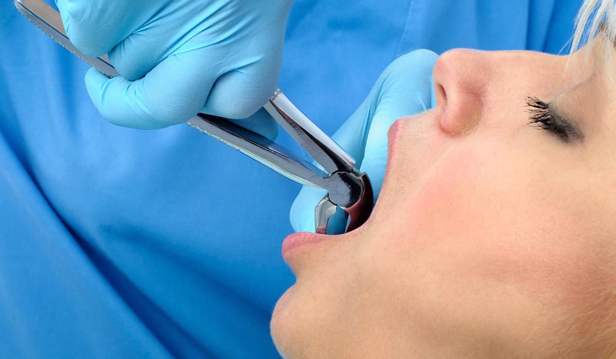
Wisdom teeth are permanent molars that usually emerge during the late teens or early twenties. It’s not uncommon for these teeth to be associated with pain or complications. Dentists often recommend having the wisdom teeth removed. Any tooth removal can be concerning for divers since keeping a scuba regulator in place means securing it with your bite.

“Wisdom teeth” is the common name for the third molars. There are typically four wisdom teeth. They are the farthest back tooth on each side of the upper and lower jaws, and their location and large size make them perfect for grinding food. They are vestigial teeth from our hominid ancestors who had to grind tough, fibrous vegetables. Genetic variation in jaw and tooth size, a diet less stimulating to jaw growth and lack of tooth loss contribute to a lack of space for the wisdom teeth. In a healthy mouth with enough space, the third molars may not need removal.

Wisdom Tooth Complications

The physical eruption of wisdom teeth can cause discomfort. Erupted third molars are often malformed and can be associated with weakened soft tissue. They are difficult to clean, which also makes them susceptible to decay and soft tissue infections.

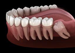
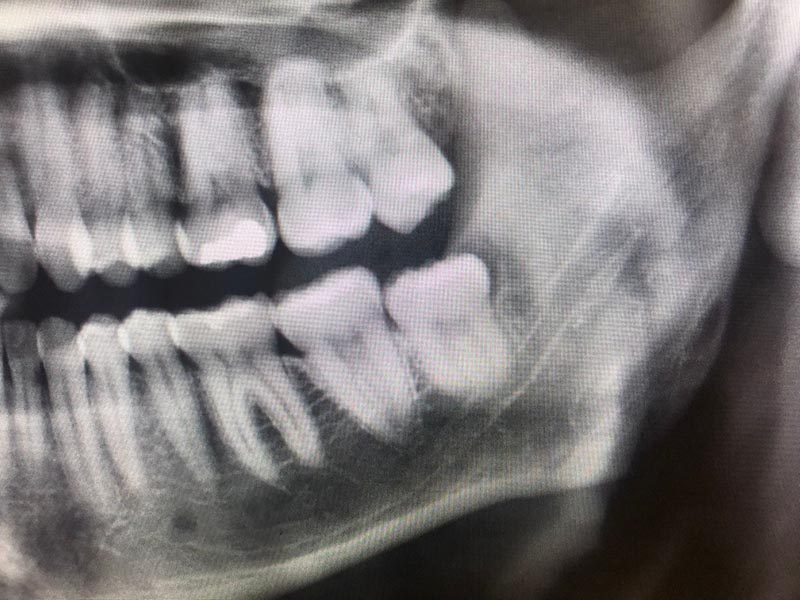
Approximately one-third of all wisdom teeth do not fully erupt into position. This lack of eruption is called impaction. Some third molars remain deep within the jaw; others are only slightly malpositioned or have a small flap of soft tissue overlying part of the tooth. Impaction impedes proper cleaning, which can lead to a buildup of bacteria that may cause an infection. This infection, called pericoronitis, typically involves the soft tissue around the crown of a partially erupted third molar. Treatment of pericoronitis consists of oral antibiotics, chlorhexidine rinses and warm saline rinses. Problems with a wisdom tooth can lead to decay and infection in the adjacent second molar.

An infected third molar will usually cause pain and swelling. As the infection progresses, swelling increases and can cause trismus (locked jaw) or limited jaw opening. Some infections are minor while others may be life-threatening. One dangerous complication is when the position of an infected lower third molar lets the infection extend into deep spaces in the neck or affect the airway.

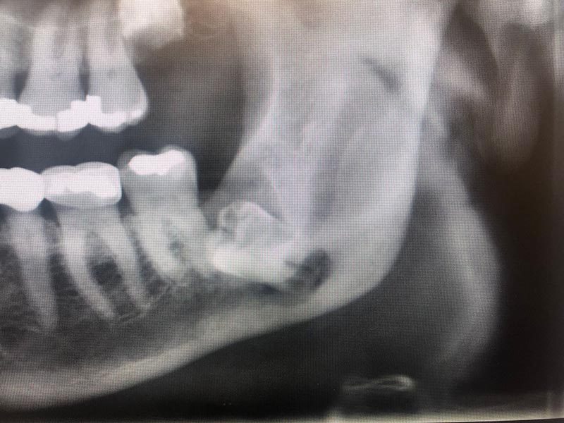
Impacted teeth can be associated with cysts and tumors, but the most common problem is infection. Less frequently, associated cysts can become infected or grow large enough to weaken the lower jaw. A weakened jaw can fracture with minimal trauma.

Complications From Extraction

Among the possible complications following wisdom tooth removal are dry socket, extraction-site infection and acute maxillary sinusitis.

  • Dry socket is a painful condition resulting from the loss of a blood clot in the extraction site. The standard treatment is oral antibiotics and medicated dressing changes.
  • Infection of the extraction site can occur immediately after the extraction and continue to be a risk for about a month. It is treated with oral antibiotic therapy and often a drainage procedure.
  • The maxillary third molar is usually close to the maxillary sinus and can affect the thin bone between the tooth and the sinus. This can result in infection and, rarely, an opening from the sinus into the mouth. An uncomplicated acute maxillary sinusitis usually responds to antibiotic therapy. A chronic opening or chronic sinusitis might require surgical intervention.
Note proximity of third molar root to the maxillary sinus. (Image courtesy Nicolas S. Veaco, D.D.S., M.D., M.S.)
The removal of some deeply impacted teeth can decrease the integrity of the mandible, putting it at risk for fracture. (Image courtesy Nicolas S. Veaco, D.D.S., M.D., M.S.)

Implications in Diving

Plan any dental surgical procedures well ahead of a dive trip, especially a trip to a remote location. There are four primary circumstances in which wisdom teeth may affect diving.

  • Tooth removal before diving: There is a possibility of unrecognized bleeding from the extraction site and the flow of compressed gas into the oral cavity. Avoid diving immediately after dental extraction, even if there were no complications.
  • Problems during a trip: Dental care can be challenging in a remote location; a cavity, toothache, soft tissue infection or tooth squeeze has the potential to disrupt a dive vacation. Most of the time you can avoid these complications with adequate prevention. If you have not had your wisdom teeth removed you may wish to inquire about the need to remove them. Maintain your dental health as part of remaining fit to dive.
  • Diving after removal of an impacted wisdom tooth: Ask for advice from the surgeon who removed the tooth or teeth. Numbness, as well as dental and muscle pain, may impair your ability to hold a mouthpiece in place. A loosely held regulator can be a drowning hazard. Most specialists recommend a minimum of four weeks after an uncomplicated wisdom tooth surgery. Even after that, complications are possible. Deeply impacted wisdom teeth that result in nerve damage, sinus complications or weakening of the lower jaw might require months to heal.
  • Jaw softness: This is much less common. Impacted teeth can be associated with large cysts or chronic infection, which can weaken the lower jaw or interfere with standard drainage of the maxillary sinuses. When this happens, the end of the jaw remains more fragile while healing. Such an issue would appear during a routine examination with appropriate imaging. Returning to dive too early could increase the risk of fracture.

Other Considerations for Divers

  • If there is still some localized edema (swelling), off-gassing of nitrogen from the area during decompression may be impaired. Although decompression illness in a small area of the jaw seems unlikely, this could be a problem.
  • Some types of pain medicine could pose a risk during dives. Drugs like codeine, oxycodone and other narcotics could synergize with the narcotic effect of inert gases (nitrogen narcosis), dangerously impairing performance and judgment underwater.
  • If you still have symptoms after the extraction procedure, you should avoid diving until you are symptom-free.

Guidelines for Diving After Dental Surgery

  • Wait for a minimum of four to six weeks or until the tooth socket and/or oral tissue has healed sufficiently to minimize the risk of infection or further trauma.
  • Don’t dive until you have discontinued using medication to control pain resulting from the surgery. Thus you will avoid any risk of drug interaction with nitrogen.
  • Make sure you can hold the regulator mouthpiece without pain or discomfort for long enough to perform a planned dive.
  • For more information on dental health, see the Mayo Clinic’s guide.

Special thanks to Nicolas S. Veaco, D.D.S., M.D., M.S. for his contributions and revisions

Box Jellyfish

Box jellyfish (cubozoans) are cube-shaped medusa notorious for having one of the most potent venoms known. Certain species can kill an adult human in as little as three minutes, scarcely enough time for any rescue response.

Medusas are the migrant forms of cnidarians. In the case of box jellyfish, their belllike bodies are cube shaped with tentacles extending from each corner. Box jellyfish are complex animals with a propulsion mechanism and a relatively sophisticated nervous system for a jellyfish. They have up to 24 eyes, some of them with corneas and retinas, enabling them to not only detect light but also to see and circumnavigate objects to avoid collision. While some jellyfish live off of symbiotic algae, box jellyfish prey on small fish, which are immediately paralyzed upon contact with their tentacles. The tentacles are then retracted, carrying the prey into the bell for digestion. Some species hunt during the day and can at night be observed resting on the ocean floor.

Mechanisms of Injury

From 1884 to 1996 there were more than 60 reported fatalities from box jellyfish stings in Australia. There are species of box jellyfish in almost all tropical and subtropical seas, but life-threatening species seem to be restricted to the Indo-Pacific.

Here are some notorious species to be aware of.

  • Sea Wasp (Chironex fleckerii): Found in the coastal waters of Australia and Southeast Asia, the sea wasp is the common name for the most dangerous cnidarian. The scientific name of this monster is Chironex fleckerii. C. fleckerii is the largest species of cubozoan. Sea wasps have a bell approximately 8 inches (20 centimeters) in diameter and tentacles ranging from a few centimeters to up to 10 feet (3 meters). Contact with these animals triggers the most powerful and lethal envenomation process known to science. Sea wasp envenomation causes immediate excruciating pain followed by cardiac failure. Death may occur in as little as three minutes. Recent studies have identified a component of the venom that drills a hole in red blood cells, causing a massive release of potassium, possibly responsible for the lethal cardiovascular depression. The same study may have also identified a way to inhibit this effect, which in the coming years could prove to be clinically promising.
  • Four-Handed Box Jellyfish (Chiropsalmus quadrumanus): The four-handed box jellyfish ranges from South Carolina to the Caribbean, the Gulf of Mexico and as far south as Brazil. The four-handed box jellyfish can inflict extremely painful stings and is the slightly smaller American cousin to the Australian sea wasp. There is one documented case of a four-year-old boy who was stung in the Gulf of Mexico and died within 40 minutes.
  • Bonaire Banded Box Jellyfish (Tamoya ohboya): Tamoya ohboya is a newly discovered, highly venomous species found in the Dutch Caribbean. Since 1989 there have been roughly 50 confirmed sightings, primarily in Bonaire, with the remainder on the shores of Mexico, St. Lucia, Honduras, St. Vincent and the Grenadines. There have only been three reported cases of envenomation, which led to intense pain and skin damage; only one case required hospitalization.
  • Carukia Barnesi & Malo Kingi: Tiny box jellyfish found near Australia, Carukia barnesi and Malo kingi, are responsible for the infamous and extremely painful symptomatic complex known as Irukandji syndrome. These small cubozoans’ bells measure only a few millimeters and their tentacles may be as long as 3 feet (1 meter). Fortunately, fatalities from these smaller species are rare, but stings are extremely painful and can cause systemic symptoms including cardiovascular instability that should prompt immediate medical attention. Survivors have reported a feeling of impending doom, claiming they were certain that they could not survive such intense, generalized pain; however, it is important to note that a single sting should not be fatal. Though stings from lesser-known species of cubozoans are not necessarily lethal, they can still be very painful. An immediate medical evaluation is always recommended.

Prevention

  • Properly research the areas where you intend to dive or swim. Some jellyfish are seasonal or nocturnal.
  • Avoid known box jellyfish habitats if you are not sure the dive site or swimming area is safe. If you are stung, cardiovascular stability can rapidly deteriorate with very little time for any effective field intervention.
  • In northern Queensland, Australia, net enclosures are placed in the water where box jellyfish are known to be during summer months (November to May), but these cannot guarantee safety.
  • Minimize unprotected areas. Wear full exposure suits, hoods, boots and gloves. Something as simple as nylon pantyhose worn over the skin can prevent jellyfish stings.
  • Carry sufficient household vinegar with you to all dive sites.

First Aid

If stung by any jellyfish, follow these procedures in this order:

  • Contact local emergency medical services.
  • Avoid rubbing the area. Jellyfish tentacles can be cylindrical or flattened, but they are coated with stinging cells (cnidocytes). Rubbing the area before removing any remaining tentacles will cause these tentacles to roll over the skin, significantly increasing the affected surface area and the envenomation process.
  • Monitor the victim’s circulation, airway and breathing. Be prepared to perform CPR at any moment (particularly if you suspect a box jellyfish sting).
  • Apply household vinegar to the area. Generously pour or spray the area with vinegar for no less than 30 seconds to neutralize any invisible remnants. You can pour the vinegar over the area or use a spray bottle, which optimizes application. Let the vinegar stand for a few minutes before doing anything else. This will not do anything to the pain or the venom already injected, but it is intended to stabilize any remaining unfired nematocysts on the diver’s skin before you try to remove them.
  • Wash the area with seawater (or saline). Use a syringe with a steady stream of water to help remove any tentacle remains. Do not rub. Do not use fresh water; this could cause and worsen the discharge from the tentacle.
  • Apply heat. Immerse the affected area in hot water (no hotter than 113°F/45°C) for 30 to 90 minutes. If you are assisting a sting victim, test the water on yourself first to assess the heat level. Do not rely on the victim’s assessment, as intense pain may impair their ability to evaluate tolerable heat levels. If you cannot measure water temperature, a good rule of thumb is to use the hottest water you can tolerate without scalding. Remember that different body areas have different tolerance to heat, so test the water on yourself on the same area where the diver was injured. Repeat if necessary. If hot water is not available, apply a cold pack or ice in a dry plastic bag. Application of heat has two purposes: 1) It may mask the perception of pain, and 2) it may assist in breaking down the venom molecules. Since we know the venom is a protein that has been superficially inoculated, application of heat may help by denaturing the toxin.
  • Always seek an emergency medical evaluation.

For additional information about marine life injuries, check out the Hazardous Marine Life Medical Reference Book.

Hot Tubs After Diving

Getting into a hot tub immediately after diving alters decompression stress. Hot tubs could cause a positive or negative response depending on the magnitude of the inert gas load and the heat stress.

Temperature and Heat Exchange

Our bodies maintain a state of internal dynamic stability. While many biological processes are occurring, the net result is a relative balance of forces and reactions. This is called homeostasis, and temperature regulation (thermoregulation) is an important aspect.

Physiological responses to changes in external temperature promotes thermal equilibrium. Cold stress causes blood vessels in the periphery (limbs and surface tissues) to shrink (vasoconstriction). This reduces peripheral blood flow and makes the outer shell of the body more of a passive thermal barrier to help protect core temperature. Substantial cold stress can also promote shivering, with the potential to increase the metabolic rate five to seven times that of rest. Shivering is effective in warming the body.

Heat stress causes the same blood vessels to dilate (vasodilation), increasing blood flow through the outer shell. A warmer shell increases radiative heat loss if the surrounding temperature is lower than skin temperature. The combination of a high skin temperature and an increased sweat rate enhances heat loss through the evaporation of moisture on the skin in air. Evaporation will not occur in a wet environment, but substantial heat energy can be transferred to the water through contact (conduction) some through convection as current replace warmed water with cooler air to be heated.

Peripheral Temperature and Inert Gas Exchange

Vasodilation of peripheral tissues increases blood flow and inert gas uptake during the descent and bottom phases of a dive. Peripheral vasoconstriction during these phases reduces inert gas uptake. Peripheral cooling during the ascent and stop phase will impair inert gas elimination. Mild warming during ascent can promote inert gas elimination, but only effectively when modest. Rapid warming will promote bubble formation. Mild warming might occur when a diver crosses a thermocline into warmer surface water. Rapid warming can be induced by electrically heated garments turned up too quickly during the ascent or post-dive by the hot tub or hot shower.

Temperature and Bubble Formation

The solubility of gas in tissues decreases as the temperature rises. Rapidly warming tissues with high inert gas loads can promote bubble formation that can produce symptoms of decompression sickness before an increase in blood flow can remove the gas. Symptoms of skin bends are most likely, but more serious symptoms can also develop.

Dive Computer and Thermal Information

Dive computers typically measure water temperature, but none measure the thermal status of the diver. Thermal status can vary dramatically based on the protective equipment worn and the physical activity of the diver. Even if the thermal status was known, no decompression models currently incorporates this information into decompression computations in a meaningful way. 

Post-Dive Practice

Divers are responsible to consider the impact of their dive and post-dive activity. Active warming during dives increases inert gas uptake. Being cold at the end of a dive traps inert gas. Jumping into a hot tub (or hot shower) immediately post-dive increases the decompression stress and, if gas loads are substantial, the risk of decompression sickness. It may not be easy to hear, but delayed gratification is the best approach for safety. You can think about the hot tub or shower while you eat, take care of your equipment, fill out a logbook, etc. Allowing time for offgassing, especially when it is impaired by your cool state, reduces risk. The more severe the decompression stress, the longer you should wait. If you need the post-dive warming immediately post-dive you should moderate your dive profile to reduce the risk.

Recommendations

Delay the hot tub or hot shower. The longer the delay the better, particularly with high gas loads. Think of this as a surface interval, with the hot tub or shower safest as the next dive.

If you are unwilling to wait, dive more conservatively.

Use a lukewarm shower temperature if you can’t wait.

Neal W. Pollock, Ph.D.

Patent Foramen Ovale (PFO)

Patent foramen ovale (PFO) is a common congenital heart defect. It is a hole between the right and left sides of the heart. The foramen ovale is the wall separating the atria. Prior to birth, the foramen ovale has an opening that lets blood pass from the right to the left atrium. Shortly after birth this opening fuses. In about 27 percent of people, it fails to fuse completely.

With a patent foramen ovale, if the pressure in the right atrium rises above the pressure in the left atrium, blood can flow from the right to the left atrium (known as a right-to-left shunt or RLS). In divers, this may let gas bubbles from the venous blood (venous gas emboli or VGE) pass to the arterial side and cause decompression illness (DCI).

Patent Foramen Ovale and DCI Risk

Epidemiological studies show an association between PFO and certain types of neurological and cutaneous decompression illness (DCI). The risk of DCI is 2.5 times greater in divers with a PFO (and the risk of neurological DCI is 4 times greater).

The overall risk of neurological DCI is still low, even in a person with a PFO. For some people, PFO is riskier than predicted. The goals of guidelines for PFO testing are to identify such individuals and manage their DCI risk.

Testing for Patent Foramen Ovale

Routine screening for patent foramen ovale during a dive medical fitness assessment (either initial or periodic) is not necessary. You should have a PFO test if you have a history of more than one episode of DCI with cerebral, spinal, eighth cranial nerve or cutaneous manifestations.

Neither noncutaneous manifestations of mild DCI nor isolated headaches after diving are indications for PFO screening.

  • The testing must include bubble contrast, ideally combined with a transthoracic echocardiogram (TTE). The use of two-dimensional and color-flow echocardiography without bubble contrast is not adequate.
  • The testing must attempt to provoke right-to-left shunting, including Valsalva release or sniffing, while the right atrium is densely opaque with bubble contrast.
  • A spontaneous shunt without provocation or a significant provoked right-to-left shunt with VGE following diving is a risk factor for DCI with cerebral, spinal, vestibulocochlear or cutaneous manifestations.
  • Smaller shunts are associated with a lower but poorly defined risk of DCI. The significance of minor degrees of RLS needs evaluation in the clinical setting that led to testing.
  • Detection of a PFO after an episode of DCI does not guarantee that the PFO contributed.

Positive PFO Test

Your diagnosis may be a PFO that is likely to increase DCI risk. If so, you can consider the following options in consultation with a diving physician:

  • Stop diving.
  • Dive more conservatively. Various strategies may reduce the risk of significant venous bubble formation after diving or the subsequent right-to-left shunting of bubbles across a PFO. It is best to discuss with a dive medicine expert the appropriateness of this approach and the strategies you can use. These strategies may include:
    • Reducing dive times to well inside accepted no-decompression limits
    • Doing only one dive per day
    • Planning for air dives but breathing nitrox (while abiding by the actual gas’s maximum operating depth)
    • Lengthening a safety stop or your decompression time at shallow stops
    • Avoiding heavy exercise and unnecessary lifting or straining for at least three hours after diving
  • Close the PFO. Closing a PFO after an episode of DCI does not ensure that DCI will not occur again. All of the options require careful consideration of the risks and benefits and the clinical setting that led to screening.

Return to Diving After Closure

Following the closure of a PFO and before returning to diving, you should have a repeat bubble-contrast echocardiogram. This test should demonstrate shunt closure at a minimum of three months after the surgery. You should only resume diving once a test confirms closure of your PFO and you are no longer taking potent antiplatelet medication.

Note: Venous bubbles can also enter systemic circulation through intrapulmonary shunts, although the role of this pathway in DCI is less well understood than that of a PFO. These shunts are typically closed at rest. They tend to open with exercise, hypoxia and beta-adrenergic stimulation and close with hyperoxia. Any of these happening after a dive could cause DCI when it might not otherwise have occurred; administration of supplemental oxygen is likely to minimize this effect.

Divers With PFO

  • The estimated incidence of neurological DCI in divers with PFO is 4.7 DCI cases per 10,000 dives.
  • Four studies showed that the prevalence of RLS or a large PFO in divers with spinal DCI is 44% compared to 14.2% in those without either RLS or PFO.
  • Half of the divers in the studies with RLS-related DCI have a PFO that is a centimeter in diameter or larger. The highest risk of DCI is in those with the largest PFOs.
  • Cerebral, spinal, cutaneous and inner ear DCI are all associated with PFO. The link between PFO and cutaneous and inner ear DCS is the strongest. In approximately 74% of cases with inner ear symptoms and no other symptoms of hyperbaric-related issues, 80% of the cases had a large, spontaneously shunting PFO.
  • For a PFO to contribute to DCI, you need to have a large PFO, VGE must form, something must cause the PFO to open, bubbles must cross it, and the bubbles must reach a target tissue while it is still supersaturated and vulnerable.

For more information about PFO, visit Risk Mitigation for Divers With a Known PFO.

Pacemakers

Pacemakers are small, battery-operated devices that help the heart beat in a regular rhythm. They do this by generating a mild electrical current that stimulates the heart to beat.

The device is implanted under the skin of the chest just below the collarbone. It connects to the heart with tiny wires that thread into the heart through the major blood vessels. In some individuals, the heart may need only intermittent help from the pacemaker if the pause between two beats becomes too long. In others, however, the heart may depend entirely on the pacemaker for regular stimulation.

Fitness to dive

An Implantable Cardioverter Defibrillator or ICD pacemaker with leads and modem for telemonitoring at home. The device sends data to the hospital on a regular basis.

A pacemaker experiences the same ambient pressures as the diver. For recreational diving, a suitable pacemaker must perform at a maximum depth of at least 130 feet (40 meters). It must operate satisfactorily during conditions of relatively rapid pressure changes on ascent and descent.

A pacemaker and cardiac defibrillator are not the same — both may be needed by some patients. The need for a cardiac defibrillator is a contraindication for diving. It is implanted for those at high risk for cardiac arrest and shock if indicated. A period of altered consciousness and subsequent drowning is likely during this period of defibrillator intervention while underwater.

As with any medication or medical device, the underlying problem that led to the implantation of the pacemaker is the most significant factor in determining someone’s fitness to dive. Every case involving a pacemaker is specific to the individual.

The two most important factors to consider are the following:

Implications in Diving

The need for a pacemaker usually indicates a disturbance in the heart’s conduction system. The disturbance might be from structural damage to the heart muscle, as is often the case following a major heart attack. In this case one might lack the necessary cardiovascular fitness to dive safely.

You might depend on a pacemaker because the area that generates the impulses that make the heart muscle contract does not function consistently or adequately. The circuitry that conducts the impulses to the heart muscle may be faulty, resulting in improper or irregular signals. Without the assistance of a pacemaker, one might suffer episodes of loss of consciousness or fainting.

Due to the risk of drowning, any medical condition that could predispose a diver to suffer a sudden loss of consciousness is a contraindication for diving.

A mild heart attack could cause minimal residual damage to your heart muscle. Even with minimal damage, the conduction system could be unreliable and dependent upon a boost from a pacemaker. A cardiologist can determine if your level of cardiovascular fitness is sufficient for safe diving. Your pacemaker needs to be rated to function at a pressure of at least 130 feet (40 meters) to be considered fit for recreational diving.

Any divers with heart issues should have an evaluation for medical fitness before diving. A significant number of recreational diving fatalities each year are attributable to coronary artery disease.

For the Diver

  • Diving often occurs in remote locations far from facilities that provide an adequate level of cardiac care for someone with a pacemaker.
  • Be mindful of your cardiovascular condition and the remoteness of your dive locations.

For the Dive Operator

  • Consider each individual’s health status on a case-by-case basis. Divers or dive students with significant cardiovascular disease and less-than-optimal exercise tolerance should be discouraged from participating in scuba diving. The individual must have adequate exercise tolerance for safe diving under routine conditions. They must also possess enough cardiovascular reserve to perform at the higher level needed in emergencies.
  • Assessing someone’s cardiovascular fitness to dive is beyond the scope of a dive instructor. When in doubt, encourage your client to present a medical clearance to scuba dive signed by their physician and keep a copy for your records.
  • If you are in doubt or feel uncomfortable about your client’s safety, remember you always have the right of refusal.
  • Resource: Right of Refusal – Alert Diver © Q4 Fall 2019.
  1. Why is the individual dependent on a pacemaker? The most common reason people depend on pacemakers is underlying ischemic heart disease. Although the pacemaker might address the cardiac dysrhythmia, the underlying heart disease might still be significant enough to disqualify someone from diving.
  2. Is the individual’s pacemaker rated to perform as intended at depths under the pressures involved in recreational diving plus an added margin of safety? Although there are no air pockets in these devices, and there are no reports of case deformation due to pressure, this concern is still valid. BCD straps over the device might cause discomfort.

Portuguese Man-of-War (Bluebottle)

Portuguese man-of-wars are free-floating cnidarians with blue gas-filled bladders and long tentacles that drift on the surface of the ocean. Contact with a man-of-war’s tentacles can cause intense pain and other, systemic symptoms.

There are two species in the genus: Physalia physalis (known as Portuguese man-of-war) in the Atlantic, and Physalia utriculus (known as bluebottle) in the Indo-Pacific. These animals are easily recognized; if you see a blue, purple or pinkish gas-filled bladder with blueish tentacles, you can bet it is a specimen of Physalia.

Although a Portuguese man-of-war appears to be a single creature, it is actually a colony made up of four types of polyps.

The Atlantic (P. physalis) species is typically a bit larger than its Australian counterpart (P. utriculus), with the gas bladder (pneumatophore) rarely exceeding a foot (30 centimeters) and its tentacles (dactylozoids) commonly 33 feet (10 meters) long and possibly extending up to 100 feet (30 meters).

Although many people think these animals are a species of jellyfish, Portuguese man-of-war belong to the order Siphonophora, a class of hydrozoans. Despite their resemblance to jellyfish, these animals are more closely related to fire coral and stinging hydroids than to true jellyfish.

Mechanisms of Injury

These animals contain cnidocytes capable of delivering a potent proteic neurotoxin capable of paralyzing small fish.

Approximately 10,000 people are stung by cnidarians each summer off the coasts of Australia, and the vast majority of these stings are by Physalia species. In fact, man-of-wars cause the most cnidarian envenomations leading to emergency evaluations globally. The risk may not be so great for divers, however, as most Physalia stings occur on beaches or on the surface of the water rather than while submerged. Certain regions are known to have seasonal outbreaks, but incidence is highly variable between regions.

Signs and Symptoms

Physalia stings cause red welts accompanied by swelling and moderate to severe pain. These local symptoms last for two to three days. Systemic symptoms are less frequent but potentially severe. They may include generalized malaise, vomiting, fever, an elevated heart rate at rest (tachycardia), shortness of breath and muscular cramps in the abdomen and back. Severe allergic reactions to the man-of-war’s venom may interfere with cardiac and respiratory function, so divers should always seek a prompt professional medical evaluation.

Prevention

  • Always look up and around while surfacing. Pay special attention during the last 15 to 20 feet of your ascent, since this is the area where you are most likely to encounter cnidarians and their submerged tentacles.
  • Wear full-body exposure suits regardless of water temperature. Mechanical protection is the best way to prevent stings and rashes. Even thin rash guards or dive skins are usually sufficient to prevent direct contact with most cnidarians.
  • In areas where these animals are known to be endemic, a hooded vest may be the best way to protect your face, ears and neck.

First Aid

  • Avoid rubbing the area.
    • Cnidarian tentacles are like nematocyst-coated spaghetti, so rubbing the area or allowing the tentacles to roll over unaffected skin will significantly increase the affected surface area and the envenomation process. NOTE: Initial pain may be intense. Though life-threatening complications are rare, monitor circulation, airway and breathing, and be prepared to perform CPR if necessary.
  • Remove the tentacles.
    • Carefully remove the man-of-war’s tentacles to avoid further envenomation using tweezers or gloves. NOTE: If you do not have access to tweezers or gloves, the keratin layer of the skin on your fingers is likely thick enough to protect you. Keep in mind, however, that after removal your fingers may contain hundreds or thousands of unfired nematocysts, so pretend you have been handling hot chili peppers that could cause painful blisters anywhere you touch; so treat your fingers according to the following instructions.
  • Flush the area with seawater.
    • Once the tentacles and any remnants have been removed, use a high-volume syringe to flush the area with a powerful stream of seawater. The purpose of this is to remove any remaining unfired nematocysts. Never use fresh water since this will cause unfired nematocysts to fire (via osmotic lysis).
  • Apply vinegar.
    • White household vinegar (or a mild acetic-acid solution of 2 to 5 percent in water) tends to stabilize unfired nematocysts. Vinegar will not do anything to the venom already injected, so it is only used as a rescue technique to prevent further envenomation from unfired nematocysts.
  • Apply heat.
    The application of heat has two purposes: 1) It may mask the perception of pain, and 2) it may assist in thermolysis. Since we know the venom is a protein that has been micro-inoculated superficially, application of heat may help by denaturing these toxins.
    • Immerse the affected area in hot water (no hotter than 113°F/45°C) for 30 to 90 minutes. If you are assisting a sting victim, test the water on yourself first to assess heat level. Do not rely on the victim’s assessment of a hot but tolerable temperature, as intense pain may impair their ability to evaluate safe heat levels. If you cannot measure water temperature, a good rule of thumb is to use the hottest water you can tolerate without scalding. Note that different body areas have different tolerance to heat, so test the water on yourself on the same area where the diver was injured.
    • Repeat as necessary.
    • If hot water is not available, apply a cold pack or ice in a dry plastic bag.
  • Always seek a professional medical evaluation.
  • Continue monitoring.
    • Continue monitoring the diver until a higher level of care has been reached. Injuries involving species of Physalia tend to cause intense systemic symptoms. Shock might result from substantial envenomations or in smaller victims.

Vinegar and Physalia Species

Use of vinegar is controversial with Physalia spp. Though the use of vinegar has traditionally been recommended, several studies both in-vivo and in-vitro show massive nematocyst discharge upon pouring vinegar over certain species of cnidarians, including Physalia. Still, the most current American Heart Association guidelines (AHA 2015) recommend application of vinegar for all jellyfish — including Physalia spp.

If you choose to apply vinegar, you can optimize application and reduce waste by using spray bottles. Generously spray the area with vinegar for no less than 30 seconds to neutralize invisible remnants. Pick off any remaining tentacles.

Implications in Diving

For the Diver

For the Dive Operator

For the Physician

  • Administer first aid treatment as described above.
  • Seek a professional medical evaluation. Any doctor should be able to help, regardless of any dive medicine knowledge or training.
  • A return to diving may be considered if a physician determines that the injury is closed and there are no unacceptable risks of infection.
  • As the leader of the expedition, you have a duty of care if the injured person was hurt during your trip.
  • Provide first aid treatment as descried above.
    • There are many folkloric first aid treatments proposals; use common sense, and refrain from attempting any scientifically unsound solutions. Remember you might be liable.
  • Make sure you have your customers evaluated by a medical professional.
  • Don’t worry about finding a doctor with dive medicine experience; any doctor should be able to help.
  • Treatment is usually symptomatic, with an emphasis on pain management. Do not hesitate to manage the symptoms aggressively. Pain can be significant to extreme. Shock is possible with significant envenomations and in small victims. Physalia spp tend to cause systemic symptoms beyond those expected at the area of contact.
  • Corticosteroids may or may not be effective.
  • Venoms from these hydrozoans are known to have dermonecrotic and hemolytic effects. Monitor accordingly.

For additional information about marine life injuries, check out the Hazardous Marine Life Medical Reference Book.

Cone Snails

Cone snails are marine gastropods characterized by a conical shell and beautiful color patterns. Cone snails possess a harpoonlike tooth capable of injecting a potent neurotoxin that can be dangerous to humans.

There are about 600 species of cone snails, all of which are poisonous. Cone snails live in shallow reefs partially buried under sandy sediment, rocks or coral in tropical and subtropical waters. Some species have adapted to colder waters.

Mechanisms of Injury

Injuries typically occur when the animal is handled. Cone snails administer stings by extending a long flexible tube called a proboscis and then firing a venomous, harpoonlike tooth (radula).

Signs and Symptoms

A cone snail sting can cause mild to moderate pain, and the area may develop other signs of an acute inflammatory reaction such as redness and swelling. Conus toxins affect the nervous system and are capable of causing paralysis, which may lead to respiratory failure and death.

Prevention

If you see a beautiful cone-shaped marine snail, it is probably a cone snail. It’s difficult to tell whether a cone snail is inhabiting a given shell, as they are able to hide deep inside them. Since all cone snails are venomous, err on the side of safety, and do not touch it.

First Aid

Unfortunately there is no specific treatment for cone-snail envenomations. First aid focuses on controlling pain but may not influence outcomes. Envenomation will not necessarily be fatal, but depending on the species, the amount of venom injected, and the victim’s size and susceptibility, complete paralysis may occur, and this may lead to death. Cone snail venom is a mixture of many different substances, including tetrodotoxin (TTX).

The prevalence and incidence of cone snail envenomations are unknown but probably rare in divers and the general population. Shell collectors (professional or amateur) may be at higher risk.

  • Clean the wound with fresh water, and provide care for the small puncture wound.
  • Apply a pressure immobilization bandage. Application of heat might help with pain management, but since TTX is a heat-stable toxin, the application of heat will not denature the toxin.
  • Watch for signs and symptoms of progressive paralysis.
    • Be prepared to provide mechanical ventilations with a bag valve mask or a manually triggered ventilator.
    • Do not wait for signs and symptoms of paralysis. Always seek an evaluation at the nearest emergency department. The bite site might be painless and still be lethally toxic.

Implications in Diving

For the Diver

  • If you see a marine snail with a cone-shaped shell, it is best to assume it is a species of cone snail and refrain from handling it — even if the shell appears to be empty.
  • Remember all species of cone snails can cause envenomation.
  • If you or someone you are diving with has been stung, seek professional medical evaluation. Any doctor should be able to help, regardless of any dive medicine knowledge or training.
  • Do not neglect these injuries. Although fatalities are rare, they are possible.

For the Diver Operator

Strongly discourage your customers from handling these creatures or their shells.

  • As the leader of the expedition, you have a duty of care if a diver gets injured during your trip.
  • Provide first aid treatment as descried above.
    • There are many folkloric first-aid treatments proposals; use common sense, and refrain from attempting any scientifically unsound solutions. Remember you might be liable.
  • Make sure you have your customers evaluated by a medical professional.

For the Physician

  • Treatment is symptomatic.
  • Complications include flaccid paralysis.
    • Observation for 6 to 8 hours is recommended to rule out respiratory depression.
  • Conus toxins are composed of several proteins, carbohydrates and quaternary ammonium derivates, which can cause cardiotoxicity , convulstant activity, vasoactive effects and flaccid paralysis.
  • Thermocoagulation could help denature some toxin components at the wound site.
  • There is no antivenom available.

For additional information about marine life injuries, check out the Hazardous Marine Life Medical Reference Book.

O’Neill Grading System

The O’Neill grading system is a way to categorize the severity of middle-ear barotrauma (MEBT). It is simple and is intended to provide consistency in diagnosis with sufficient details to direct treatment.

Grade 0: Eustachian Tube Dysfunction

  • Baseline photo depicting anatomical appearance of the tympanic membrane (TM) before exposure to pressure
  • Symptoms with no anatomical change (no trauma) from baseline

Grade 1 Barotrauma

  • Erythema increased from baseline
  • Fluid or air trapping (visible bubble) in the middle-ear space

Grade 2 Barotrauma

  • Any bleeding noted within the tympanic membrane or middle-ear space
  • Perforation

Blue-Ringed Octopus

Blue-ringed octopuses are small, venomous octopuses that live in tropical tide pools from southern Japan to the coastal reefs of Australia and the western Indo-Pacific. These small octopuses are the only cephalopods known to be dangerous to humans.

The blue-ringed octopus hardly ever exceeds 8 inches (20 centimeters) in size. Their most distinctive feature is the blue iridescent rings that cover their yellow-colored body; however, it is important to note that this feature is only displayed when the animal is disturbed, hunting or mating. When calm or at rest, the animal may display an overall yellowish, grey or beige coloration without any visible blue rings. The blue-ringed octopus is more active at night, spending most of the day hidden in its nest in shallow areas or tide pools.

There are three species of blue-ringed octopuses:

  • Greater blue-ringed octopus (Hapalochlaena lunulata)
  • Southern (or lesser) blue-ringed octopus (Hapalochlaena maculosa)
  • Blue-lined octopus (Hapalochlaena fasciata)

Blue-ringed octopus envenomations are very rare. Cases outside of southern Japan, Australia and the western Indo-Pacific are generally due to deliberate handling of aquarium specimens. There have been only a handful of reported fatal cases. Full recovery can be expected with timely professional medical intervention.

Mechanism of Injury

Like all cephalopods, octopuses have strong beaks similar to those of parrots and parakeets. All octopuses have some sort of venom to paralyze their victims, but the blue-ringed octopus bite may contain an extremely powerful neurotoxin called tetrodotoxin (TTX), which can be up to 10,000 times more potent than cyanide and can paralyze a victim in minutes. Theoretically, a little more than one-half milligram of this venom — an amount that could fit on the head of a pin — is enough to kill an adult human. Certain bacteria present in the blue-ringed octopus’ salivary glands synthesize the toxin. TTX is not unique to the blue-ringed octopus; certain newts, dart frogs, cone snails and pufferfish can also be a source of TTX intoxication, though by different mechanisms.

Signs and Symptoms

A blue-ringed octopus bite is usually painless or no more painful than a bee sting; however, even painless bites should be taken seriously. Neurological symptoms dominate every stage of envenomation and manifest as paresthesia (tingling and numbness) progressing to paralysis that could potentially culminate in death. If envenomation has occurred, signs and symptoms usually start within minutes and may include paresthesia of the lips and tongue. This is usually followed by excessive salivation, trouble with pronunciation (dysarthria), difficulty swallowing (dysphagia), sweating, dizziness and headache. Serious cases may progress to muscular weakness, loss of coordination, tremors, and paralysis. Paralysis may eventually affect respiratory muscles, which can lead to severe hypoxia with cyanosis (blue or purple tissue discoloration due to insufficient oxygen in the blood).

First Stage: Initial Presentation

  • Prickling and tingling (paresthesia) of the lips and tongue, followed by facial and extremity tingling and numbness
  • Headache, sensations of lightness or floating
  • Profuse sweating (diaphoresis)
  • Dizziness
  • Salivation (ptyalism)
  • Nausea, vomiting (emesis), diarrhea, abdominal (epigastric) pain
  • Difficulty moving (motor dysfunction)
  • Weakness (malaise)
  • Speech difficulties

Second Stage

  • Progressive paralysis
    • First in the extremities
    • Then in the rest of the body
    • Finally in the respiratory muscles; causing
      • difficulty breathing or shortness of breath (dyspnea)
      • abnormal heart rhythms (cardiac dysrhythmias or arrhythmia)
      • abnormally low blood pressure (hypotension)
      • fixed and dilated pupils (mydriasis)
      • coma, seizures, respiratory arrest, and death

Prevention

  • These animals are not aggressive, and divers should not fear blue-ringed octopuses.
  • If encountered, remain at arm’s length and avoid handling these animals.

Due to their small size and lack of skeleton, a blue-ringed octopus den might be a small space only accessible through a tiny crevice, so avoid picking up bottles, cans or mollusk shells in areas these octopuses are known to inhabit.

First Aid

Care is supportive. There is no antivenom available.

  • Clean the bite site with freshwater, and provide care for a small puncture wound.
  • Apply a pressure immobilization wrap. TTX is a heat-stable toxin, which means application of heat will not alter the it.
  • Watch for signs and symptoms of descending paralysis. Be prepared to provide mechanical ventilations with a bag valve mask or manually triggered ventilator. Seek professional medical care (do not wait for signs and symptoms of paralysis). Evaluation and observation should be sought at the nearest emergency department or medical facility. Beware: The bite site might be painless and still be lethally toxic.
  • Wound excision is never recommended.

Implications in Diving

For the Diver

  • Administer first aid as described above.
  • Although the bite might be almost imperceptible and painless, immediately seek professional medical evaluation. Never underestimate a bite from a blue-ringed octopus. The toxin from these animals can kill a human in as little as 20 minutes.

For the Diver Operator

  • As the leader of the expedition, you have a duty of care if the injured person was hurt during your trip.
  • Provide first aid treatment as descried above.
    • There may be many folkloric first aid treatments; use common sense, and refrain from attempting any scientifically unsound solutions. Remember you might be liable.
  • Take your customer to the closest medical facility. Never underestimate a bite from a blue-ringed octopus. The toxin from these animals can kill a human in as little as 20 minutes.
  • Don’t worry about finding a doctor with dive medicine experience; these envenomations are not the purview of a dive medicine expert but rather an emergency medicine specialist or toxicologist.

For the Physician

  • See first aid treatment described above.
  • Definitive treatment is symptomatic and supportive. All fatalities reported have been due to respiratory depression.
  • TTX poisoning may either have rapid onset (10 to 45 minutes) or delayed onset (generally within 3 to 6 hours but rarely longer).
    • Death may occur as early as 20 minutes or as late as 24 hours after exposure; it usually occurs within the first 4 to 8 hours.
    • Patients who live through the acute intoxication in the first 24 hours usually recover without residual deficits. Symptoms may last for several days, and recovery takes days.

For additional information about marine life injuries, check out the Hazardous Marine Life Medical Reference Book.

Sea Urchins

Sea urchins are typically small, round, spiny creatures found on shallow, rocky marine coastlines. The primary hazard associated with sea urchins is contact with their spines.

Sea urchins are echinoderms, a phylum of marine animals that also includes starfish, sand dollars and sea cucumbers. Echinoderms are recognizable because of their pentaradial symmetry (they have five rays of symmetry), which is easily observed in starfish. This symmetry corresponds with a water vascular system used for locomotion, transportation of nutrients and waste, and respiration. Sea urchins have tubular feet called pedicellariae, which enable movement. In one genus of sea urchin — the flower sea urchin — some of the pedicellariae have evolved into toxic claws. In these animals, the spines are short and harmless, but these toxic claws can inflict an envenomation.

Sea urchins feed on organic matter in the seabed. Their mouth is located at the base of their shell, and their anus is on the top. Sea urchins’ color varies by species — shades of black, red, brown, green, yellow and pink are common.

There are species of sea urchins in all oceans, from tropical to arctic waters. Most incidents between humans and sea urchins occur in tropical and subtropical waters. The flower sea urchin (Toxopneustes spp.) is the most toxic of all sea urchins. Its short spines are harmless, but its pedicellariae, which look like small flowers, are tiny claws (Toxopnueustes means “toxic foot”). These claws contain a toxin that can cause severe pain and other symptoms similar to those of a jellyfish sting.

Mechanism of Injury

Sea urchins are covered in spines, which can easily penetrate divers’ boots and wetsuits, puncture the skin and break off. These spines are made of calcium carbonate, the same material that makes up eggshells. Sea urchin spines are usually hollow and can be fragile, particularly when the time comes to extract broken spines from the skin.

Injuries usually happen when people step on urchins while walking across shallow rocky bottoms or tide pools. Divers and snorkelers are often injured while swimming on the surface in shallow water, as well as when entering or exiting the water from shore dives.

Although little epidemiological data is available, sea urchin puncture wounds are common among divers, particularly when in shallow waters, near rocky shores or close to wrecks or other hard surfaces.

Signs and Symptoms

Injuries generally take the form of puncture wounds, often multiple and localized. Skin scrapes and lacerations are also possible. Puncture wounds are typically painful and associated with redness and swelling. Pain ranges from mild to severe depending on several factors, including the species of sea urchin, the location of the wound on the body, whether joints or deeper muscle tissues are involved, number of punctures, depth of puncture and the individual’s pain tolerance. Multiple puncture wounds may cause limb weakness or paralysis, particularly with the long-spined species of the genus Diadema. On very rare occasions, immediate life-threatening complications may occur.

Prevention

  • Be observant while entering or exiting the water from shore dives, particularly when the bottom is rocky.
  • If swimming, snorkeling or diving in shallow water, near a rocky shore or close to wrecks or other hard surfaces, maintain a prudent distance and good buoyancy control.
  • Avoid handling these animals.

First Aid

There is no universally accepted treatment for sea urchin puncture wounds. Both first aid and definitive care are symptomatic.

  • Apply heat. Immerse the affected area in hot water (no hotter than 113°F/45°C) for 30 to 90 minutes. If you are assisting a sting victim, first test the water yourself to assess heat. Do not rely on the victim’s assessment, as pain may impair their ability to evaluate heat. If you cannot measure the water temperature, a good rule of thumb is to use the hottest water you can tolerate without scalding. Different body areas have different tolerance to heat, so test the water on yourself on the same area where the diver was injured. Repeat if necessary. 
    • Very few species of sea urchins contain venom. If venom is present, hot-water immersion may also help denature any superficial toxins.
  • Remove any superficial spines. Tweezers can be used for this purpose, but sea urchin spines are hollow and can be very fragile when grabbed from the sides. Your bare fingers are a softer alternative to tweezers. 
    • Do not attempt to remove spines embedded deep in the skin; let medical professionals handle those. Deeply embedded spines may break down into smaller pieces, complicating the removal process.
  • Wash the area thoroughly, but avoid forceful rubbing and scrubbing if you suspect there may still be spines embedded in the skin.
  • Apply an antiseptic or over-the-counter antibiotic ointment if available.
  • Do not close the wound with tape or glue; this might increase the risk of infection. 
    • Deep puncture wounds are the perfect environment to culture an infection, particularly tetanus.

Regardless of any first aid provided, always seek a professional medical evaluation.

Implications in Diving

For the Diver

  • Seek a professional medical evaluation as soon as reasonably possible. Do not neglect these wounds.
    • Don’t worry about finding a doctor with dive medicine experience; any doctor should be able to help.
  • Puncture wounds in the vicinity of a joint can be problematic.
  • Do not neglect these wounds, some of the complications could have a negative impact for life.

For the Dive Operator

  • As the leader of the expedition, you have a duty of care if the injured party was injured during your trip.
  • Provide first aid treatment as descried above.
    • There are many folkloric first aid treatments proposed for sea urchin puncture wounds; use common sense, and refrain from attempting any scientifically unsound solutions. Remember, you might be liable.
  • Have the customer evaluated by a medical professional — any doctor should be able to help.

For the Physician

  • There is no universally accepted treatment. For the most part, treatment is symptomatic.
  • Very few species of sea urchins are actually toxic.
  • Antigens present on the teguments covering the spines could be causing swelling.
  • The decision of whether surgical removal of retained spines is necessary is usually based on joint or muscular layer involvement and whether there is pain with movement or signs of infection.
  • Remaining spines will usually encapsulate in a short time, but they may not always dissolve.
  • Although these wounds don’t always get infected, it is worth considering prophylactic antibiotic therapy.
  • Make sure your patient has adequate immunization for tetanus. Deep puncture wounds are potentially tetanogenic.
  • Reassess frequently over the first few days to monitor progress and possible infection.

For additional information about marine life injuries, check out the Hazardous Marine Life Medical Reference Book.