Arrhythmias/Dysrhythmias

The term arrhythmia (or, sometimes, dysrhythmia) means an abnormal heartbeat. It is used to describe a number of very well defined electrophysiological manifestations ranging from benign, harmless conditions to severe, life-threatening disturbances of the heart’s rhythm.

The heart’s electrical wiring is one of the most sophisticated and enduring pieces of nature’s engineering. It controls the rate at which your heart has been beating, every minute, hour and day, 365 days a year since before you were born. Such a precise function on such a vital organ, disease or damage to that wiring can cause symptoms and increase the risk of premature death.

Divers should be aware of its potential implications in diving; and any physicians who treat them should be familiar with their effects on the safety of scuba divers.

Symptoms

A normal heart beats between 60 to 100 times per minute. In well-trained athletes, or even some non-athletic individuals, a heart at rest may beat at as slowly as 40 to 50 times a minute. Entirely healthy, normal individuals experience occasional irregular isolated heart beats or minor changes in their heart’s rhythm. These isolated extra heart beats can be caused by drugs (such as caffeine), or stress, or can occur for no apparent reason.

Dysrhythmias become serious only when they are prolonged or when they do not result in proper and effective contraction of the heart. Physiologically significant extra heartbeats (also known as extrasystoles) may originate in the upper chambers of the heart (this is called supraventricular tachycardia) or in the lower chambers of the heart (this is called ventricular tachycardia). The cause of these extra beats may be a short circuit, an additional parallel conduction pathway in the heart’s wiring, or it may be the result of some other more complex cardiac disorder. People who have episodes or periods of rapid heartbeat are at risk of losing consciousness during such events. Other people have a fairly stable dysrhythmia (such as fixed atrial fibrillation) but in conjunction with additional cardiovascular disorders or other health problems that may exacerbate the effect of their rhythm disturbance. An abnormally slow heartbeat (or a heart blockage) may cause symptoms as well as concerns.

Implications in Diving

The risk for any person who develops a dysrhythmia during a dive is loss of consciousness; which exposes the diver to leading to an unacceptably high risk of drowning. Serious dysrhythmias, such as ventricular tachycardia (VT) and many types of atrial dysrhythmias, are incompatible with diving. Supraventricular tachycardia, for example, is unpredictable in its onset and may even be triggered simply by immersing one’s face in cold water. Anyone who has had more than one episode of this type of dysrhythmia should not dive.

Most dysrhythmias that require medication to be controlled often disqualify the affected individual from safe diving. Exceptions may be made on a case-by-case basis in consultation with a cardiologist and a diving medical officer.

An individual who has any cardiac dysrhythmia needs a complete medical evaluation by a cardiologist prior to engaging in any diving activities.

Electrophysiology studies (EPS) can often identify abnormal conduction pathways, and in some cases the problem can be corrected. Recently, clinicians and researchers have determined that people with some dysrhythmias (such as certain types of Wolff-Parkinson-White syndrome, which is characterized by an extra electrical pathway) may safely participate in diving after a thorough evaluation by a cardiologist. Also in select cases, people with stable atrial dysrhythmias (such as uncomplicated atrial fibrillation) may dive safely if a cardiologist determines that they have no other significant health problems.

Common Dysrhythmias & Impacts on Diving

Atrial Fibrillation

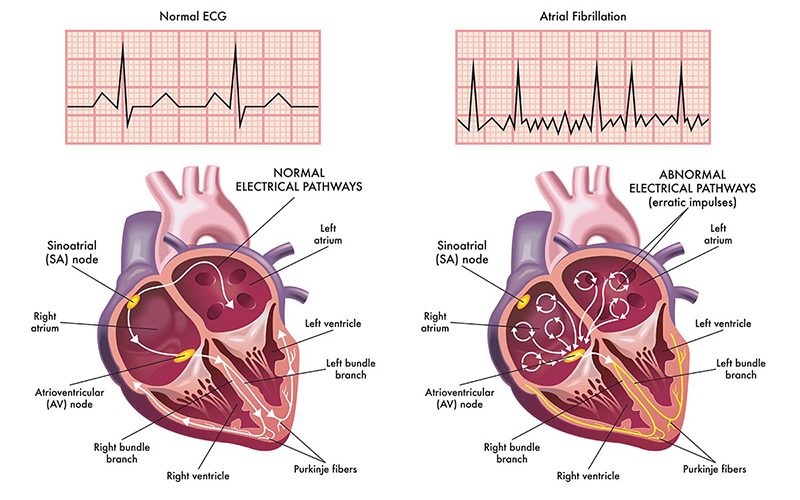
Atrial fibrillation (AF, or also known simply as AFib) is the most common form of dysrhythmia, and it is characterized by an irregular and fast heartbeat.

AF results from a disturbance of the electrical signals that normally make the heart contract in a controlled rhythm, where one can easily predict when the next heart beat will come. During an AFib, the heart’s rhythm is chaotic, and rapid impulses cause an uncoordinated atrial filling and ventricle pumping action. This leads to a decrease in overall cardiac output, which can affect one’s exercise capacity or even result in unconsciousness. In addition, AF causes blood to pool in the atria. Blood pooling promotes the formation of clots, which can break loose and enter the circulatory system. This increase the risk of a stroke, as any of these lose clots could easily impacts the brain an cause infarction.

The most common causes of AF are hypertension and coronary artery disease. Additional causes include a history of valvular disorders, hypertrophic cardiomyopathy (a thickening of the heart’s muscle), deep vein thrombosis (DVT), pulmonary embolism, obesity, hyperthyroidism, heavy alcohol consumption, an imbalance of electrolytes in the blood, cardiac surgery and heart failure.

Some people with AF experience no symptoms and are unaware they have the condition until it’s discovered during a physical examination. Others may experience symptoms such as the following:

  • Palpitations (a racing, uncomfortable, irregular heartbeat or a flip-flopping sensation in the chest)
  • Weakness
  • Reduced ability to exercise
  • Fatigue
  • Lightheadedness
  • Dizziness
  • Confusion
  • Shortness of breath
  • Chest pain

The occurrence and duration of atrial fibrillation usually falls into one of three patterns:

  • Occasional (or paroxysmal): The rhythm disturbance and its symptoms come and go, lasting for a few minutes to a few hours, and then stop on their own. Such events may occur a couple of times a year, and their frequency typically increases over time.
  • Persistent: The heart’s rhythm doesn’t go back to normal on its own, and treatment — such as an electrical shock or medication — is required to restore a normal rhythm.
  • Permanent: The heart’s rhythm can’t be restored to normal. Treatment may be required to control the heart rate, and medication may be prescribed to prevent the formation of blood clots.

Any new case of AF should be investigated and its cause determined. An investigation may include a physical exam; an electrocardiogram; a measurement of electrolyte levels, including magnesium; a thyroid-hormone test; an echocardiogram; a complete blood count; and/or a chest X-ray.

Treating the underlying cause of AF can help control the fibrillation. Various medications, including beta blockers, may help regulate the heart rate. A procedure known as cardioversion — which can be performed with either a mild electrical shock or medication — may prompt the heart to revert to a normal rhythm; before cardioversion is attempted, it is essential to ensure that a clot has not formed in the atrium. Cardiac ablation may also be used to treat AF. In addition, anticoagulant drugs are often prescribed for individuals with AF to prevent the formation clots and thus reduce their risk of stroke. It is also of note that the neurological effects of an embolic stroke associated with AF can sometimes be confused with the symptoms of decompression sickness.

Implications in Diving: A thorough medical examination should be conducted to identify the underlying cause of the atrial fibrillation. It is often the underlying cause that is of most concern regarding fitness to dive. But even atrial fibrillation itself can have a significant impact on cardiac output and therefore on maximum exercise capacity. Individuals who experience recurrent episodes of symptomatic AF should refrain from further diving. The medications often used to control atrial fibrillation can present their own problems, by causing other dysrhythmias and/or impairing the individual’s exercise capacity. It is essential that anyone diagnosed with AF have a detailed discussion with a cardiologist before resuming diving.

Extrasystoles

Heart beats that occur outside the heart’s regular rhythm are known as extrasystoles. They often arise in the ventricles, in which case they are referred to as premature ventricular contractions or sometimes premature ventricular complexes, abbreviated as PVCs. The cause of such extra beats is often benign, but it can also result from serious underlying heart disease.

PVCs are common even in healthy individuals; they have been recorded in 75 percent of those who undergo prolonged cardiac monitoring (for at least 24 hours, that is). The incidence of PVCs also increases with age; they have been recorded in more than 5 percent of individuals more than 40 years old who undergo an electrocardiogram (or ECG, a test that typically takes less than 10 minutes to administer). Men seem to be affected more than women.

The extrasystole itself is usually not felt. It is followed by a pause — a skipped beat — as the heart’s electrical system resets itself. The contraction following the pause is usually more forceful than normal, and this beat is frequently perceived as a palpitation — an unusually rapid or intense beat. If extrasystoles are either sustained or combined with other rhythm abnormalities, affected individuals may also experience dizziness or lightheadedness. Heart palpitations and the sensation of missed or skipped beats are the most common complaints of those who seek medical care for extrasystole.

A medical examination of the condition begins with a history and physical, and should also include an ECG and various laboratory tests, including the levels of electrolytes (such as sodium, potassium and chloride) in the blood. In some cases, doctors may recommend an echocardiogram (an ultrasound examination of the heart), a stress test and/or the use of a Holter monitor (a device that records the heart’s electrical activity continuously for a 24- to 48-hour period). Holter monitoring may uncover PVCs that are unifocal — that is, they originate from a single location. Of greater concern are multifocal PVCs — those that arise from multiple locations — as well as those that exhibit specific patterns known R-on-T phenomenon, bigeminy and trigeminy.

If serious structural disorders, such as coronary artery disease or cardiomyopathy (a weakening of the heart muscle), can be ruled out — and the patient remains asymptomatic — the only “treatment” required may be reassurance. But for symptomatic patients, the course is less clear, as there is controversy regarding the effectiveness of the available treatment options. Two drugs commonly used to treat high blood pressure — beta blockers and calcium channel blockers — have been used in patients with extrasystole with some success. Antiarrhythmics have also been prescribed for extrasystole but have met with mixed reviews. A procedure known as cardiac ablation may be an option for symptomatic patients, if the location where their extra beats arise can be identified; the procedure involves threading tiny electrodes into the heart via catheters, then zapping the affected locations to rewire the heart’s faulty circuits.

Implications in Diving: Although PVCs are present in a large percentage of otherwise normal individuals, they have been shown to increase mortality over time. If PVCs are detected, it is important that they be investigated and that known associated conditions be ruled out. Divers who experience PVCs and who are found to also have coronary artery disease or cardiomyopathy will put themselves at significant risk if they continue to dive. Divers diagnosed with R-on-T phenomenon, nonsustained runs of ventricular tachycardia or multifocal PVCs should likewise refrain from diving. Divers who experience PVCs but remain asymptomatic may be able to consider a return to diving; such individuals should discuss with their cardiologist their medical findings, their desire to continue diving and their clear understanding of the risks involved.

Hazardous Marine Life Injuries

During any dive, you may encounter a less-than-friendly undersea critter — which could result in a serious skin reaction or injury. How you respond to the injury could impact symptoms and the overall healing process.

If you are taking medications to treat a sting or wound, in general, it is safe to dive while taking an antibiotic or corticosteroid medication. If a wound infection is more than minor or is expanding, however, diving should be curtailed until it becomes minor, is no longer progressing and/or can be easily covered with a dressing. In or out of the water, corticosteroid medication should always be taken with the understanding that a rare side effect is to cause serious deterioration of the head (“ball” of the ball-and-socket joint) of the femur, the long bone of the thigh.

Most injuries from animals result from chance encounters. Be an alert diver, and respect their personal space. From coral scrapes to envenomations by various marine life, here are recommendations to treat certain marine life injuries.

Coral Scrapes

Coral scrapes are among the most common injuries from marine life incurred by divers and snorkelers. The surface of coral is covered by soft living material, which is easily torn from the rigid (abrasive) structure underneath, and thus deposited into the scrape or cut. This greatly prolongs the wound-healing process by causing inflammation and, occasionally, initiating an infection. Cuts and scrapes from sharp-edged coral and barnacles tend to fester and may take weeks or even months to heal.

Treatment

  1. Scrub the cut vigorously with soap and water, and then flush the wound with large amounts of water.
  2. Flush the wound with a half-strength solution of hydrogen peroxide in water. Rinse again with water.
  3. Apply a thin layer of bacitracin, mupirocin (Bactroban) or other similar antiseptic ointment, and cover the wound with a dry, sterile, and non-adherent dressing. If no ointment or dressing is available, the wound can be left open. Thereafter, it should be cleaned and re-dressed twice a day. If the wound develops a pus-laden crust, you may use “wet-to-dry” dressing changes to remove the upper non-healing layer in order to expose healthy, healing tissue. This is done by putting a dry sterile gauze pad over the wound (without any underlying ointment), soaking the gauze pad with saline or a dilute antiseptic solution (such as 1- to 5-percent povidone-iodine in disinfected water), allowing the liquid to dry, and then ripping the bandage off the wound. The dead and dying tissue adheres to the gauze and is lifted free. This method may be painful for the patient. The pink (hopefully), slightly bleeding tissue underneath should be healthy and healing. Dressings are changed once or twice a day. Wet-to-dry dressings are used for a few days, until they become non-adherent or the tissue appears infection-free. At that point, switch back to No. 3 above.
  4. If the wound shows any sign of infection (extreme redness, pus, swollen lymph glands), the injured person (particularly one with impairment of his or her immune system) should be started by a qualified health professional on an antibiotic, taking into consideration the possibility of a Vibrio infection. Vibrio bacteria are found more often in the marine environment than on land, and can rapidly cause an overwhelming illness and even death in a human with an impaired immune system (e.g., someone with AIDS, diabetes or chronic liver disease). A common oral antibiotic that is usually effective against Vibrio species is ciprofloxacin (Cipro).

Coral poisoning occurs if coral abrasions or cuts are extensive or are from a particularly toxic species. Symptoms include a wound that heals poorly or continues to drain pus or cloudy fluid, swelling around the cut, swollen lymph glands, fever, chills and fatigue. If these symptoms are present, the injured person should see a physician, who may elect to treat the person with an antibiotic and/or corticosteroid medication.

Sea Urchin Spine Punctures

Some sea urchins are covered with sharp venom-filled spines that can easily penetrate and break off into the skin. Others (found in the South Pacific) may have small pincer-like appendages that grasp their victims and inoculate them with venom from a sac within each pincer. Sea urchin punctures or stings are painful wounds, most often of the hands or feet. If a person receives many wounds simultaneously, the reaction may be so severe as to cause extreme muscle spasm, difficulty in breathing, weakness and collapse.

Treatment

  1. Immerse the wound in non-scalding hot water to tolerance (110 to 113° F/43.3 to 45° C). This frequently provides pain relief. Other field remedies, such as application of vinegar or urine, are less likely to diminish the pain. If necessary, administer pain medication appropriate to control the pain.
  2. Carefully remove any readily visible spines. Do not dig around in the skin to try to fish them out – this risks crushing the spines and making them more difficult to remove. Do not intentionally crush the spines. Purple or black markings in the skin immediately after a sea urchin encounter do not necessarily indicate the presence of a retained spine fragment. The discoloration more likely is dye leached from the surface of a spine, commonly from a black urchin (Diadema species). The dye will be absorbed over 24 to 48 hours, and the discoloration will disappear. If there are still black markings after 48 to 72 hours, then a spine fragment is likely present.
  3. If the sting is caused by a species with pincer organs, use hot water immersion, then apply shaving cream or a soap paste and shave the area.
  4. Seek the care of a physician if spines are retained in the hand or foot, or near a joint. They may need to be removed surgically, to minimize infection, inflammation and damage to nerves or important blood vessels.
  5. If the wound shows any sign of infection (extreme redness, pus, swollen regional lymph glands) or if a spine has penetrated deeply into a joint, the injured person should be started by a qualified health professional on an antibiotic, taking into consideration the possibility of a Vibrio infection (see No. 4 under Coral Scrapes).
  6. If a spine puncture in the palm of the hand results in a persistent swollen finger(s) without any sign of infection (fever, redness, swollen lymph glands in the elbow or armpit), then it may become necessary to treat the injured person with a seven- to 14-day course of a non-steroidal anti-inflammatory drug (e.g., ibuprofen) or, in a more severe case, with oral prednisone, a corticosteroid medication.

Lionfish, Scorpionfish & Stonefish Envenomations

Lionfish (as well as scorpionfish and stonefish) possess dorsal, anal and pelvic spines that transport venom from venom glands into puncture wounds. Common reactions include redness or blanching, swelling, and blistering. The injuries can be extraordinarily painful and occasionally life-threatening (in the case of a stonefish).

Treatment

Soaking the wound in non-scalding hot water to tolerance (110 to 113° F/43.3 to 45° C):

  • may provide dramatic relief of pain from a lionfish sting,
  • is less likely to be effective for a scorpionfish sting, and
  • may have little or no effect on the pain from a stonefish sting, but it should be done nonetheless, because the heat may inactivate some of the harmful components of the venom.

If the injured person appears intoxicated or is exhibiting signs of weakness, vomiting, short of breath or unconscious, seek immediate advanced medical care.

Wound care is standard, so, for the blistering wound noted above, appropriate therapy would be a topical antiseptic (such as silver sulfadiazene [Silvadene] cream or bacitracin ointment) and daily dressing changes. A scorpionfish sting frequently requires weeks to months to heal, and therefore requires the attention of a physician. There is an antivenin available to physicians to help manage the sting of the dreaded stonefish.

Stingray Envenomations

A stingray does its damage by lashing upward in defense with a muscular tail-like appendage, which carries up to four sharp, swordlike stingers. The stingers are supplied with venom, so that the injury created is both a deep puncture or laceration and an envenomation.

The pain from a stingray wound can be excruciating and accompanied by bleeding, weakness, vomiting, headache, fainting, shortness of breath, paralysis, collapse and occasionally, death. Most wounds involve the feet and legs, as unwary waders and swimmers tread upon the creatures hidden in the sand.

Treatment

  1. Rinse the wound with whatever clean water is available. Immediately immerse the wound in non-scalding hot water to tolerance (110 to 113° F/43.3 to 45° C). This may provide some pain relief. Generally, it is necessary to soak the wound for 30 to 90 minutes in the hot water, but take care not to create a burn wound. Gently extract any obvious piece(s) of stinger.
  2. Scrub the wound with soap and water. Do not try to sew or tape it closed – doing so could promote a serious infection by “sealing in” harmful bacteria.
  3. Apply a dressing and seek medical help. If more than 12 hours will pass before a doctor can be reached, start the injured person on an antibiotic (ciprofloxacin, trimethoprim-sulfamethoxazole or doxycycline) to oppose Vibrio bacteria.
  4. Administer pain medication sufficient to control the pain.

Prevention of Stingray Injuries

  • Always shuffle your feet when wading in stingray-infested waters.
  • Always inspect the bottom before resting a limb in the sand.
  • Never handle a stingray unless you know what you are doing or unless the stingrays are definitely familiar with divers and swimmers (e.g., the rays in “Stingray City” off Grand Cayman Island in the British West Indies). Even then, respect them for the wild creatures they are — the less you handle them, the better for them and for you, too.

Seabather’s Eruption

Often misnamed “sea lice” (which are true crustacean parasites of fish, and which inflict miniscule bites), seabather’s eruption occurs in sea water and involves predominately bathing suit-covered areas of the skin, rather than exposed areas. The skin rash distribution is very similar to that from seaweed dermatitis (read below), but no seaweed is found on the skin.

The cause is stings from the nematocysts (stinging cells) of the larval forms of certain anemones, such as Linuche unguiculata, and from thimble jellyfishes. The injured person may notice a tingling sensation under the bathing suit (breasts, groin, cuffs of wetsuits) while still in the water, which is made much worse if he/she takes a freshwater rinse (shower) while still wearing the suit. The rash usually consists of red bumps, which may become dense and confluent. Itching is severe and may become painful.

Treatment

Treatment consists of immediate (for decontamination) application of vinegar or rubbing alcohol, although the relief may be minimal. Some persons note that topical papain (e.g., unseasoned meat tenderizer) and simultaneous brisk rubbing are effective. Others have noted relief from concentrated citrus (e.g., lime) juice applied to the skin. Topical calamine lotion with 1 percent menthol may be soothing. After decontamination, hydrocortisone lotion 1 percent twice a day may be minimally effective. More potent topical steroid preparations or oral prednisone may be prescribed by a physician to provide sufficient anti-inflammatory effect to quell the reaction somewhat. However, it is not uncommon for a patient to be miserable for a few days to two weeks.

If the reaction is severe, the injured person may suffer from headache, fever, chills, weakness, vomiting, itchy eyes and burning on urination, and should be treated with oral prednisone.

The stinging cells may remain in the bathing suit even after it dries, so once a person has sustained seabather’s eruption, the clothing should undergo machine washing or be thoroughly rinsed in alcohol or vinegar, then be washed by hand with soap and water.

Seaweed Dermatitis

Seabather’s eruption is easy to confuse with seaweed dermatitis. There are more than 3,000 species of algae, which range in size from 1 micron to 100 meters in length. The blue-green algae, Microcoleus lyngbyaceus, is a fine, hair-like plant that is found in the waters near Hawaii and Florida, and gets inside the bathing suit. Out of water, the skin under the suit remains in moist contact with the algae (the other skin dries or is rinsed off), and becomes red and itchy, with occasional blistering and/or weeping. The reaction may start a few minutes to a few hours after the victim leaves the water.

The Treatment

Treatment consists of a vigorous soap-and-water scrub, followed by a rinse with isopropyl (rubbing) alcohol. Apply 1 percent hydrocortisone lotion twice a day. If the reaction is severe, oral prednisone may be administered.

Swimmer’s Itch

Also called “clamdigger’s itch,” swimmer’s itch is caused by skin contact with cercariae, which are the immature larval forms of parasitic schistosomes (flatworms) found throughout the world in both fresh and salt waters. Snails and birds are the intermediate hosts for the flatworms. They release hundreds of fork-tailed microscopic cercariae into the water.

The affliction is contracted when a film of cercariae-infested water dries on exposed (uncovered by clothing) skin. The cercariae penetrate the outer layer of the skin, where itching is noted within minutes. Shortly afterwards, the skin becomes reddened and swollen, with an intense rash and, occasionally, hives. Blisters may develop over the next 24 to 48 hours.

Untreated, the affliction is limited to 1 to 2 weeks. Persons who have suffered swimmer’s itch previously may be more severely affected on repeated exposures, which suggests that an allergic response may be a factor.

The Treatment

Swimmer’s itch can be prevented by briskly rubbing the skin with a towel immediately after leaving the water, to prevent the cercariae from having time to penetrate the skin. Once the reaction has occurred, the skin should be lightly rinsed with isopropyl (rubbing) alcohol and then coated with calamine lotion. If the reaction is severe, the injured person may be treated with oral prednisone.

Because the cercariae are present in greatest concentration in shallow, warmer water (where the snails are), swimmers should try to avoid these areas.

Jellyfish Stings

“Jellyfish” is the term commonly used to describe an enormous number of marine animals that are capable of inflicting a painful, and occasionally life-threatening, sting. These include fire coral, hydroids, jellyfishes (including “sea wasps“) and anemones. The stings occur when the victim comes into contact with the creature’s tentacles or other appendages, which may carry millions of small stinging cells, each equipped with venom and a microscopic stinger.

Depending on the species, size, geographic location, time of year and other natural factors, stings can range in severity from mild burning and skin redness to excruciating pain and severe blistering with generalized illness (nausea, vomiting, shortness of breath, muscle spasm and low blood pressure). Broken-off tentacles that are fragmented in the surf or washed up on the beach can retain their toxicity for months and should not be handled, even if they appear to be dried out and withered.

The box jellyfish (Chironex fleckeri) of northern Australia contains one of the most potent animal venoms known to man. A sting from one of these creatures can induce death in minutes from cessation of breathing, abnormal heart rhythms and very low blood pressure (shock).

Treatment

Be prepared to treat an allergic reaction following a jellyfish sting. If possible, carry an allergy kit, including injectable epinephrine (adrenaline) and an oral antihistamine.

The following therapy is recommended for all unidentified jellyfish and other creatures with stinging cells:

  1. If the sting is believed to be from the box jellyfish (Chironex fleckeri), immediately flood the wound with vinegar (5 percent acetic acid). Keep the injured person as still as possible. Continuously apply the vinegar until the individual can be brought to medical attention. If you are out at sea or on an isolated beach, allow the vinegar to soak the tentacles or stung skin for 10 minutes before attempting to remove adherent tentacles or to further treat the wound. In Australia, surf lifesavers (lifeguards) may carry antivenin, which is given as an intramuscular injection.
  2. For all other stings, if a topical decontaminant (e.g., vinegar, isopropyl [rubbing] alcohol, one-quarter-strength household ammonia, or baking soda) is available, apply it liberally onto the skin. If it is a liquid, continuously soak a compress. (Be advised that some authorities advise against the use of alcohol because of scientific evaluations that have revealed that some nematocysts discharge because of this chemical’s application.) Since not all jellyfish are identical, it is extremely helpful to know ahead of time what works for the stingers in your specific geographic location. Apply the decontaminant for 30 minutes or until pain is relieved. A paste made from unseasoned meat tenderizer (do not exceed 15 minutes’ application time, particularly upon the sensitive skin of small children) or papaya fruit may be helpful. Concentrated citrus (e.g., lime) juice may be helpful. Do not apply any organic solvent, such as kerosene, turpentine or gasoline. Until the decontaminant is available, you may rinse the skin with sea water. Do not simply rinse the skin gently with fresh water or apply ice directly to the skin. A brisk freshwater stream (forceful shower) may have sufficient force to physically remove the microscopic stinging cells, but non-forceful application is more likely to cause the cells to fire, increasing the envenomation. A non-moist ice or cold pack may be useful to diminish pain, but take care to wipe away any surface moisture (condensation) prior to the application.
  3. After decontamination, apply a lather of shaving cream or soap and shave the affected area with a razor. In a pinch, you can use a paste of sand or mud in sea water and a clamshell.
  4. Reapply the primary decontaminant for 15 minutes.
  5. Apply a thin coating of hydrocortisone lotion (0.5 to 1 percent) twice a day. Anesthetic ointment (such as lidocaine hydrochloride 2.5 percent or a benzocaine-containing spray) may provide short-term pain relief.
  6. If the victim has a large area involved (entire arm or leg, face, or genitals), is very young or very old, or shows signs of generalized illness (nausea, vomiting, weakness, shortness of breath or chest pain), seek help from a doctor. If a person has placed tentacle fragments in his mouth, have him swish and spit whatever potable liquid is available. If there is already swelling in the mouth (muffled voice, difficulty swallowing, enlarged tongue and lips), do not give anything by mouth, protect the airway and rapidly transport the victim to a hospital.

Ciguatera Poisoning

Ciguatera fish poisoning involves a large number of tropical and semitropical bottom-feeding fish that dine on plants or smaller fish, which have accumulated toxins from microscopic dinoflagellates, such as Gambierdiscus toxicus. Therefore, the larger the fish, the greater the toxicity. The ciguatoxin-carrying fish most commonly ingested include the jack, barracuda, grouper and snapper.

Symptoms, which usually begin 15 to 30 minutes after eating the contaminated fish, include abdominal pain, nausea, vomiting, diarrhea, tongue and throat numbness, tooth pain, difficulty in walking, blurred vision, skin rash, itching, tearing of the eyes, weakness, twitching muscles, incoordination, difficulty sleeping and occasional difficulty in breathing. A classic sign of ciguatera intoxication is the reversal of hot and cold sensations (hot liquids seem cold and vice versa), which may reflect general hypersensitivity to temperature.

Persons can become severely ill shortly after they are poisoned, with heart problems, low blood pressure, deficiencies of the central and peripheral nervous systems, and generalized collapse. Unfortunately, many of the debilitating, but not life-threatening, symptoms may persist in varying severity for weeks to months.

Treatment

Treatment is for the most part based upon symptoms without any specific antidote, although certain drugs are beginning to prove useful for aspects of the syndrome, such as intravenous mannitol for abnormal nervous system behavior and abnormal heart rhythms. A physician must undertake these therapies.

Prochlorperazine may be useful for vomiting; hydroxyzine or cool showers may be useful for itching. There are chemical tests to determine the presence of ciguatoxins in fish and in the bloodstream of humans, but not yet a specific antidote. If a person displays symptoms of ciguatera fish poisoning, they should be see a physician promptly.

During recovery from ciguatera poisoning, the affected person should exclude the following from their regular diet: fish, fish sauces, shellfish, shellfish sauces, alcoholic beverages, nuts and nut oils.

Paul S. Auerbach, M.D., M.S.

Seabather’s Eruption

Seabather’s eruption is lesions found primarily on parts of the body covered by swimwear. The lesions may also appear on your armpits, neck and occasionally on your arms and legs. Most divers and swimmers call it “sea lice.” That term is a misnomer. Sea lice are fish parasites that do not affect humans and have nothing to do with seabather’s eruption, which is caused by the larvae of jellyfish.

This image has an empty alt attribute; its file name is rash-mishap-T49BX087-DAN-256x312-1.jpg

Mechanisms of Injury

The primary offenders in Florida and the Caribbean are the larvae of thimble jellyfish (Linuche unguiculata). These larvae, generally half a millimeter in length, can find their way into bathing suits — even passing through the mesh of some suits — where they become trapped against the skin and will sting. The larvae are visible to the naked eye, but they become nearly invisible in the water. A lack of adult thimble jellyfish in the area is no guarantee that there won’t be larvae. One of the best signs of larvae is the appearance of a rash on swimmers or divers.

The larvae are most prevalent in April through July, although they may appear at any time. The symptoms will appear very soon (24 hours or less) after exposure to the organism and will persist for several days. Some cases record a three- or four-day delay in onset and symptoms lasting several weeks.

Signs and Symptoms

  • Itchy skin eruptions with small blisters and elevated skin areas
  • Fever
  • Headache
  • Chills
  • Nausea and vomiting

Prevention

  • Wear a wetsuit or impermeable dive skin
  • Avoid t-shirts and one-piece bathing suits, which may trap the larvae
  • Remove your wetsuit, dive skin or bathing suit after diving or swimming in areas with jellyfish larvae
  • Do not shower with your wetsuit, dive skin or bathing suit on (the fresh water may discharge nematocysts)
  • Do not wear the same clothing again without washing — the larvae may remain in the fabric

Treatment

Many of the symptoms are consistent with other illnesses. Diagnosis can be difficult unless the physician knows of the diver’s exposure to contaminated water. Often the symptoms are very mild, and a physician may consider or diagnose other causes at first. Symptoms can often be severe in children, although adults have also shown severe reactions.

Cases of seabather’s eruption will often clear spontaneously, but others may require treatment. You can consider antihistamines and anti-itching agents, but these are not proven to have significant or consistent results.

Children and individuals with allergies or diseases affecting the immune system may be at risk for severe reactions. Fortunately, the severe reaction is rare, but it can be a danger for some individuals. In these cases, some doctors prefer to use cortisone by tablet or injection.

G. Yancey Mebane, M.D.

Tympanic Membrane Rupture (Perforated Eardrum)

Tympanic membrane (TM) perforation is a tear of the eardrum, which can occur while diving due to failed middle-ear equalization.

Mechanisms of Eardrum Injury

The eardrum is a tissue separating the external ear from the middle-ear space. It is attached to a chain of small bones (auditory ossicles) located in the middle ear. The eardrum also serves as a barrier between the sterile middle-ear space and the ambient environment. The pars tensa is the tightly stretched portion of the eardrum. It consists of three layers and is the main structure of the membrane. The pars flaccida is a small triangular portion of the eardrum that consists of two layers and is quite fragile. Although the pars tensa is more robust than the pars flaccida, the pars tensa is more commonly associated with perforations.

Eardrum rupture can be caused by descending without equalizing the pressure in the middle ear, a forceful Valsalva maneuver, an explosion, a blow to the ear or head, or acoustic trauma. It is usually painful; rupture relieves the pressure (and pain) in the middle ear and may result in vertigo. There may be some bleeding in the ear canal. Contributing factors include congestion, inadequate training and excessive descent rates.

Signs and Symptoms

  • Ear pain during the descent that stops suddenly, sometimes with a loud pop (this pop usually relieves the pressure and can cause pain)
  • Bubbles coming out of your ear while equalizing
  • Clear or bloody drainage from the ear
  • Hearing loss
  • Ringing in the ear (tinnitus)
  • Lightheadedness or dizziness
  • Vertigo (spinning sensation)
  • Nausea or vomiting (usually as a result of vertigo)

Prevention

  • Do not dive when congested.
  • Refrain from diving when feeling popping or crackling in your ears, or if you have a feeling of fullness in your ears after diving.
  • Learn and use proper equalization techniques.

First Aid

  • Do not use any ear drops. If ear drops reach the middle ear, they could make things worse.
  • Lie down and rest. Keep movements and physical activity to a minimum.
  • Lying down and closing your eyes may help with vertigo, which might be significant and will likely make you feel miserable. Try to remain calm. Vertigo is usually accompanied by nausea and vomiting.
  • Seek professional medical evaluation ASAP. Any doctor should be able to help, regardless of any dive medicine knowledge or training.

Implications for Diving

For the Diver

  • If you suspect you have had a tympanic membrane rupture you should stop diving immediately.
  • If you dive with a rupture, water could pass through your ear canal into the middle ear. This could cause a sudden onset of vertigo. Never attempt to continue diving with earplugs.
  • Avoid any Valsalva-like maneuvers for middle-ear equalization, sneezing and nose blowing.
  • If you are having vertigo, your inner ear might be compromised as well. Valsalva-like maneuvers might exacerbate vertigo. Do not lift heavy weights.

For the Dive Operator

  • Provide first aid treatment, as described above. As the expedition’s leader, you have a duty of care for a diver injured during your trip.
    • Be skeptical of folkloric first aid treatments. Use common sense, and don’t attempt magic solutions. Remember that you might be liable.
  • Have the diver sit down, and reassure them during the process.
  • Help them deal with vertigo, which can be a very uncomfortable feeling that will likely make the diver — and you — feel uneasy about the situation. Rapid movements of the head and Valsalva-like maneuvers (such as lifting heavy things) might exacerbate vertigo. People with vertigo usually have:
    • A spinning sensation: They feel they are spinning or that the environment is spinning around them.
    • Repetitive nystagmus: Involuntary eye movement that can occur from side to side, up and down, or in a circular motion.
    • Nausea and vomiting: Make sure the diver does not aspirate vomit.
  • Have the diver evaluated by a medical professional in a timely fashion.
  • Don’t worry about finding a doctor with dive medicine experience. An ear, nose and throat (ENT) specialist would be ideal, but any doctor should be able to help with the initial evaluation.

For the Physician

  • Most perforated eardrums will heal spontaneously within a few weeks. It may be necessary to treat nasal and sinus congestion. If the tear or hole does not heal by itself, further treatment may involve procedures to close the perforation.
    • Eardrum patch: This is an office procedure in which an ENT applies a chemical to the edges of the tear to stimulate growth and then applies a paper patch over the hole to provide a support structure for the growth of eardrum tissue.
    • Surgery: Large eardrum defects may be fixed with surgery (tympanoplasty). An ENT surgeon takes a tiny patch of your own tissue and plants it over the hole in the eardrum. This procedure is done on an outpatient basis unless medical conditions require a longer hospital stay.
  • For assessing the severity of an ear barotrauma, use the O’Neill grading system.

Fitness to Dive

If your physician feels the healing is adequate, and there is no evidence of Eustachian tube problems, you can return to diving within several months. Chronic perforations that do not heal are a contraindication to diving.