
Few words strike fear quite like “cancer,” but what exactly is it, and how might it impact decisions about medical fitness to dive?
Cancer is a broadly applied term used to describe cells that exhibit abnormal and unregulated replication, growth and activity. These aberrant cellular behaviors can arise from alterations in DNA, or they may stem from environmental factors that disrupt DNA and thus impact normal cellular functioning. When this occurs, cells can lose the ability to control growth and may even become resistant to normal programmed cell death. Tissue masses that form as a result of this unchecked proliferation are commonly known as tumors or neoplasms, which means “new growths.” If there is a genetic basis for a neoplasm, the altered DNA can be passed to daughter cells, which then proliferate and spread. Cancer’s capacity to impact health stems from these alterations in cellular activity, which interrupt or disrupt normal organ function sufficiently to cause illness.
Cancer is the second-leading cause of death in the United States. The American Cancer Society predicts that in 2013 an estimated 1.6 million people will be diagnosed.1 With statistics like this, it is likely that you or someone you know has been or will be affected.
Diagnosis
Early detection offers the greatest hope for successful treatment. Screening has been shown to improve survival in several types of cancers. Recommended health-maintenance examinations include mammograms, cervical cytology (previously known as Papanicolaou or “Pap” smears) and colonoscopies. To demonstrate the thought process and methodology of one of these screening approaches, let’s take a closer look at colon cancer, which is the second-leading cause of cancer deaths in the United States (after lung cancer).
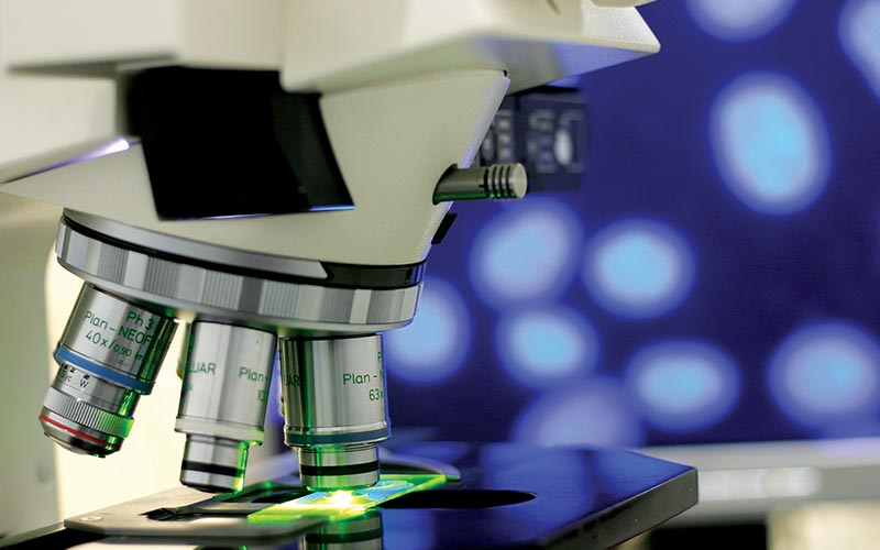
The U.S. Preventive Services Task Force recommends screening for colon cancer beginning at age 50 and continuing every 10 years until about age 75 for people of average risk.2 Colonoscopy enables detection of colon cancer as well as discovery and removal of early precancerous growths. Commonly beginning as small, benign lumps known as adenomatous polyps, these tumors may be visible in the lumen of the colon. Colonoscopy enables direct visualization of the colon’s inner surface via a camera lens connected to a flexible tube. If a polyp or other abnormal growth is detected, a tissue sample (biopsy) can be taken for further evaluation. A microscopic evaluation by a pathologist is necessary to confirm whether the biopsy is normal or neoplastic.
Typically, the first things a patient wants to know after being diagnosed with cancer is: Has it spread, and what is the prognosis (expected outcome)? To help provide a framework for such discussions and to guide treatment, early medical and surgical efforts are geared toward determining the grade and stage. A tumor’s grade is a measure of its cellular abnormality or degree of differentiation from its tissue of origin. Staging is based on the size of the primary tumor, the extent of local invasion, the spread to local lymph nodes and the presence of tumors in other areas of the body. Grade and stage are determined using techniques including clinical findings, microscopic evaluation of biopsied tissue, cytology, computerized tomography (CT), positron emission tomography (PET), magnetic resonance imaging (MRI) and surgical exploration.
Treatment
Current treatment modalities fall into four basic categories: surgery, chemotherapy, radiation and bone marrow transplantation. The specific therapeutic approach to a given cancer is influenced by many factors including size, grade and stage, location, tumor genetics (which may indicate the tumor type and sensitivity to treatment), likelihood of treatment success, risk of treatment and probability of recurrence. Treatment modalities are often provided together or in sequence to achieve remission, reduce a tumor’s impact on health or simplify surgical removal.
Benign or Malignant

Neoplasms are described as either benign or malignant — designations that are based on their potential behavior. Benign tumors generally remain localized to their site of origin, and most develop a fibrous capsule that separates them from surrounding tissue, which can make them amenable to surgical removal. While such tumors usually don’t spread from their site of origin, their size and location nonetheless can lead to significant disease if they are present in vital areas such as the brain. In this way, benign tumors that grow in confined spaces or impact other structures may be every bit as lethal as their malignant counterparts.
Malignant tumors are those most commonly referred to as cancers. Malignancy implies that the tumor can invade and destroy adjacent structures and metastasize (spread to distant sites). While not all malignant tumors behave the same, all warrant concern. The infiltrative nature of these tumors generally prompts aggressive treatment, which may include a combination of chemotherapy, radiation and surgery.
Surgery
Surgical removal of abnormal tissue is often the first-line treatment, and it can enable both gross and microscopic evaluation of tissue. Excision of wide margins of normal-appearing tissue along with areas of visible disease is frequently performed in an attempt to increase the likelihood of complete removal of the cancer cells and to minimize the risks of metastasis and local recurrence. This approach is not appropriate in every case, and its use is influenced by tumor type, location and the degree of metastasis. Tumors located in vital organs such as the brain, lungs and liver offer additional challenges for surgical resection; their removal may be life threatening or may constitute an unacceptable risk to surrounding structures. Some cancers, such as those involving blood or bone marrow, are not amenable to surgery due to the nature of the tissue. In these cases, chemotherapy and radiation are often used.
Chemotherapy
Chemotherapy refers to the use of cytotoxic drugs (drugs toxic to cells) to attempt to limit or stop tumor growth. The type of chemotherapeutic drugs used and the duration of treatment vary. Chemotherapeutic agents are delivered systemically, and many do not target tumor cells specifically; rather, they primarily affect rapidly proliferating cells.
Chemotherapy may involve one or more drugs and is often employed to kill tumor cells left behind after surgery or tumors located in inoperable locations. Chemotherapy may also be administered preoperatively when reducing tumor size will make removal safer and/or more effective. Side effects are systemic and may include fatigue, nausea, bleeding problems, diarrhea, reduced stamina, hair loss and immunosuppression. Some new agents that are more discriminating and not cytotoxic are available. An example is the use of monoclonal antibodies, which target proteins expressed in certain cancer cells.
Radiation
Radiation therapy uses focused, high-energy ionizing radiation to kill cancer cells. Radiation impacts DNA directly and/or induces formation of free radicals that alter DNA activity, leading to cell death. Because therapeutic ionizing radiation is delivered only to the site of the tumor, systemic side effects are less common than with chemotherapy. However, acute tissue damage (radiation burns) can occur, as well as delayed effects such as breakdown of local tissue or scarring that may appear months or years after treatment.
Bone Marrow Transplantation
For certain blood-based cancers, bone marrow transplantation may be used. In these cases, the patient undergoes bone marrow ablation, which kills the cancer cells along with healthy marrow. Marrow stem cells derived from either the patient or a donor are then transplanted, with the goal of growing cancer-free bone marrow.
Connection to Diving
It is important to state that diving is not associated with promoting or enhancing cancer growth.
Medical fitness to dive begins with the premise that a diver has the capacity to care for himself and assist others. Note that this starting point does not address physically disabled people, a group for whom such criteria are legitimately altered due to special procedures and methods of support. A cardinal rule of safe diving is that divers should dive within the parameters of their skills, training and physical ability. In this context, wellness encompasses the ability to handle the physical rigors of diving as well as mental and emotional readiness for the potential challenges of the environment.
With regard to medical fitness for diving with cancer, an important consideration is how the cancer affects or has the potential to affect critical organ systems in ways that may lead to injury, incapacitation or death. Factors such as fatigue, immunosuppression, bleeding and the potential need for urgent medical support require strong consideration.
Conservative postsurgical guidelines should be followed prior to a return to water sports. These include complete healing of the skin, regaining physical strength and stamina and medical clearance from the treating physician. Complete healing of wounds is critically important since both freshwater and saltwater environments are filled with bacteria that can infect open wounds.
Cancers that affect the following body systems are of particular concern to physicians who evaluate divers.
Brain
The health concerns associated with brain tumors include a potential for seizures, bleeding into the brain, sudden loss of consciousness, altered mental status, changes in behavior and diminished cognitive ability. For these reasons, recreational divers are advised to refrain from diving until their neurologist or neurosurgeon clears them. The clearance process should involve careful consideration of the diver’s likelihood for sudden neurological changes or incapacitation.
The prognosis of brain cancer is determined by the location, tumor type and extent of tissue involvement. Benign tumors that are easily amenable to surgery and located where removal involves limited damage to surrounding tissues carry a more favorable prognosis than malignant tumors that are invasive or located where removal would necessitate unacceptable risk.
Lungs
As the leading cause of cancer death, lung cancer often holds a poor prognosis. Treatment involves a variety of modalities that include chemotherapy, radiation and surgery. Tumors can cause inflammation of lung tissue, compress airways (which could predispose divers to air trapping) and decrease pulmonary function. Radiation therapy can cause lung-tissue inflammation, and surgery can cause scarring and reduce the quantity of available lung tissue. All of these issues weigh heavily in decisions about safely returning to diving and require careful consideration.
Colon

Colon cancer is the second-leading cause of cancer death, and when it’s caught late it has a poor prognosis. Treatment can include chemotherapy, radiation and surgery. Surgical resection of diseased bowel may require the creation of a colostomy or an ileostomy — a surgical opening in the abdominal wall that allows bowel contents to empty into a bag affixed externally to the skin. Such devices may be temporary or permanent.
If general health and physical endurance are consistent with safe diving practices, people can dive with ostomy bags. Special care should be taken to ensure protection of the surrounding skin, which can become irritated due to exposure to fecal contents. Starting the dive with an empty bag is also a good idea. Despite the common concern about gas expansion, no pressurized gas is added during diving, so there should not be an appreciable risk of bag rupture on ascent.
Portacaths
For patients who require repeated administration of medications or repeated blood draws, a catheter may be placed under the skin in the upper-right part of the chest. Such venous catheters are safe for divers and pose neither a risk of infection (as they are under the skin) nor of gas entry.
Conclusions
People with cancer may be medically cleared to dive, provided their health status is commensurate with strenuous activity and the cancer’s location does not place them at elevated risk of injury or loss of consciousness (lung and brain cancer are of particular concern). Divers with a recent history of cancer treatment should complete all therapy, allow sufficient time for rehabilitation and be medically cleared prior to returning to diving.
References
- American Cancer Society. Cancer Facts and Figures 2013. Atlanta: American Cancer Society; 2013. http://www.cancer.org/acs/groups/content/@epidemiologysurveilance/documents/document/acspc-036845.pdf
- Screening for Colorectal Cancer: U.S. Preventive Services Task Force Recommendation Statement 2008. http://www.uspreventiveservicestaskforce.org/uspstf08/colocancer/colors.pdf
© Alert Diver — Q2 Spring 2013